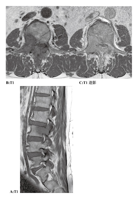
Fig. 1
Preoperative magnetic resonance imaging (MRI) of the lumbar spine. A: Midline sagittal image of the lumbar spine. The tumor expanded into the epidural space of L3. The tumor of the L3 posterior arch expanded into the paravertebral muscle. B: T1 of axial image at the L3 level. The tumor invaded the vertebral body, right pedicle, right transverse process, and spinous process of the L3 vertebra. C: gadolinium-enhanced of axial image at the L3 level. The tumor was enhanced by gadolinium.