


Chiba Medical J. 101E:1-10, 2025
doi:10.20776/S03035476-101E-1-P1
〔 Original Article 〕
Noriyasu Toshi1), Kazuhide Inage1), Kohei Okuyama1)
Geundong Kim2), Tomohito Mukaihata3), Ikuko Tajiri1)
Sumihisa Orita1,4), Yawara Eguchi1), Yasuhiro Shiga1)
Masahiro Inoue1), Miyako Suzuki-Narita5), Takahisa Hishiya1)
Takahito Arai1), Soichiro Tokeshi1), Susumu Tashiro1)
Shuhei Ohyama1), Noritaka Suzuki1), and Seiji Ohtori1)
1) Department of Orthopaedic Surgery, Graduate School of Medicine, Chiba University, Chiba 260-8670.
2) Department of Orthopaedic Surgery, Tokyo Metropolitan Bokutoh Hospital, Tokyo 130-8575.
3) Department of Orthopaedic Surgery, Katori Omigawa Medical Center, Katori 289-0332.
4) Center for Frontier Medical Engineering, Chiba University, Chiba 263-8522.
5) Department of Bioenvironmental Medicine, Graduate School of Medicine, Chiba University, Chiba 260-8670.
(Received August 21, 2024, Accepted October 21, 2024, Published March 10, 2025.)
【Introduction】The present investigation assessed the influence of romosozumab, a recently introduced medication in Japan, on the early bone fusion process in a rat posterolateral lumbar fusion (PLF) model. We aimed to overcome the limitations of a previous study by utilizing a higher dosage, which was 19 times greater than the clinical equivalent, to investigate the efficacy of lower dosages of romosozumab for promoting bone healing using the same rat lumbar PLF model.
【Methods】A total of 24 male 8-week-old Sprague–Dawley rats underwent PLF surgery and were divided into four groups according to the dosage of romosozumab administered postsurgically: the standard dose used in prior studies, half and one-tenth of this dose, and a control group. We administered romosozumab bi-weekly for 8 weeks based on the rats’ body weights. Assessments included bi-weekly micro-computed tomography (CT) scans and histological examinations of the lumbar spine performed 8 weeks post-surgery.
【Results】CT analysis revealed no significant differences in bone fusion rates and volume increases among the romosozumab groups at various time points post-surgery, but all showed significantly higher values than those in the control group. Histological evaluation confirmed these findings, with the romosozumab groups exhibiting significantly greater trabecular area ratios at the PLF site than the controls.
【Conclusions】Romosozumab effectively promotes early bone fusion at all tested dosages, including one-tenth of previously used amounts, indicating its efficacy at significantly lower doses. These findings suggest the potential clinical use of romosozumab at lower, more practical doses for enhancing bone fusion.
romosozumab, bone fusion, rat, posterolateral lumbar fusion (PLF) model
As the population ages, the likelihood of vertebral fusion procedures for those with diminished bone density is expected to rise. Complications arising from spinal fusion surgeries, such as vertebral fractures and pedicle screw loosening, are common and challenging to manage. Cadaver studies have shown a significant decrease in pullout resistance in individuals with osteoporosis, increasing the risk of pedicle screw loosening[1]. Due to the projected increase in spinal surgeries for individuals with significant bone density loss, it is crucial to prioritize rapid bone healing to minimize postoperative complications and attain optimal therapeutic outcomes.
Several pharmaceutical agents, such as bisphosphonates and synthetic parathormone derivatives, are currently utilized in the treatment of bone density loss. These therapeutic compounds influence the skeletal renewal cycle and potentially augmenting osseous resilience. The process of skeletal renewal is comprised three distinct phases. Initially, osteoclastic cells break down aged bone tissue[2]. Following this, a transitional period occurs where this degradation ceases. Lastly, osteoblastic cells generate fresh bone material. Recent scientific studies have indicated that medications aimed at preventing bone density loss may accelerate osseous repair and enhance the healing process after spinal procedures [3,4].
Romosozumab is a groundbreaking therapeutic agent that functions as a humanized immunoglobulin, promoting osteogenesis by targeting sclerostin, a crucial extracellular molecule. Sclerostin negatively regulates the canonical Wnt signaling cascade, and by disrupting its capacity to interact with low-density lipoprotein receptor-related proteins 5 and 6 (LRP5 and LRP6), romosozumab prevents the inhibition of Wnt signal propagation in osteoblastic cells, enhancing bone formation[5-7].
The Wnt signaling cascade promotes osteogenesis while decreasing osseous breakdown and enhancing mass and robustness in both compact and spongy bone tissues. Preliminary clinical trial results for romosozumab corroborate these findings. Research findings from the FRAME (Fracture study in postmenopausal women with osteoporosis) investigation showed a mean increase of 35% in the marker for bone degradation (CTX) and a statistically significant average rise of 95% in the indicator of bone production (Tracp 5b) after just one month of romosozumab therapy[6]. Additionally, our previous study confirmed its clinical effectiveness, noting significant improvements in Tracp 5b and P1NP markers and increased lumbar spine bone density at 1 month and 6 months post-administration, respectively[8].
In our department, we examined the effects of romosozumab on bone union using a rat lumbar posterior lateral fusion (PLF) model as a preliminary study. This research demonstrated early-stage bone healing and significant bone mass growth[9]. Given its effects on early healing, romosozumab appears promising for preventing postoperative complications in spinal fusion surgery. However, the prior study used a dosage of romosozumab (Evenity®) approximately 19 times higher than that typically used in clinical practice, though this was based on dosages effective in other bone healing studies[10]. To address this limitation, we aimed to investigate the efficacy of lower dosages of romosozumab for promoting bone healing using the same rat lumbar PLF model.
1.Experimental animals
The research protocol was approved by our institution’s Ethics Review Board and adhered completely to the guidelines specified in the 2011 revision of the National Institutes of Health’s Guide for the Care and Use of Laboratory Animals. The experimental methodologies employed were consistent with those used in prior investigations. For this research, we selected 24 male Sprague Dawley rodents, each approximately 8 weeks old, with body masses ranging from 200 to 250 g (Japan SLC Inc., Shizuoka, Japan).
2.Posterolateral lumbar fusion (PLF) surgery
Each rodent received an intraperitoneal injection of a mixed anesthetic solution. This solution comprised 0.15 ml/kg of medetomidine hydrochloride, 2 mg/kg of midazolam, 2.5 mg/kg of butorphanol, and 1.45 ml/kg of saline. Before the surgical procedure, a subcutaneous dose of 20,000 U/kg ampicillin sodium was administered. A midline incision was made, and the bilateral paraspinal muscle fascia was separated. This exposed the lamina, transverse processes, and intervertebral articulation of L4-L5. Approximately 40 mg of osseous tissue was extracted from the spinous processes spanning the tenth thoracic vertebra to the second lumbar vertebra for transplantation. This autologous osseous material was then bilaterally positioned between the intervertebral articulations and the transverse processes of the fourth and fifth lumbar vertebrae. All incisions were closed with 4-0 resorbable sutures. Postoperatively, the rodents were housed in cages with unrestricted access to food and water.
3.Grouping
Based on the dosage used in previous studies and accounting for body size differences, we created four groups of six rats: standard dose (25 mg/kg, R1 group), half dose (12.5 mg/kg, R1/2 group), one-tenth dose (2.5 mg/kg, R1/10 group), and a control group with normal diet (C group). The R1 group received a full dose of romosozumab (Evenity®, containing 105 mg/1.17 mL of sclerostin antibody, 2.41 mg/1.17 mL of calcium acetate hydrate [13 mM], 2.04 mg/1.17 mL of acetic anhydride [17 mM], 70 mg/1.17 mL of sucrose[6%], 0.070 mg/1.17 mL of polysorbate-20 [0.006%], at pH 5.2, Amgen Inc.). The other groups (R1/2, R1/10, and C) were administered a mixture of romosozumab (as described above) and saline (Saline, Otsuka Pharma Inc., Japan) at 12.5 mg/kg, 2.5 mg/kg, and 0 mg/kg, and at 12.5 mg/kg, 22.5 mg/kg, and 25 mg/kg, respectively. Each group was administered the respective treatments via subcutaneous injection twice a week (Tuesdays and Fridays in the morning) until the 8th week.
4.Evaluation items
Micro-computed tomography (CT) examination
Periodic assessments of the osseous transplant sites were conducted using high-resolution CT. These examinations took place at two-week intervals, starting before the surgical procedure and continuing for eight weeks post-operation. During each imaging session, the subjects were kept under controlled anesthesia with a 1.5% concentration of a volatile anesthetic agent administered via inhalation. The imaging protocol used the following technical parameters: spatial resolution of 59 μm, X-ray tube potential of 90 kV, tube current of 200 mA, a 30 mm field of view, and an exposure duration of 26 seconds. A panel of three orthopedic specialists, not involved in the research, independently assessed osseous union rates between the intervertebral articulations and transverse processes. Their evaluations were based on the acquired imaging data. To complement this qualitative assessment with quantitative metrics, specialized medical imaging software was used to measure and compare the volumetric dimensions of the fusion regions. This analysis provided objective measurements of the extent and progression of osseous integration.
Bone densitometry
Rats were euthanized by overdose of a three-agent anesthetic by the 8th week post-surgery. The right femur of each rat was disarticulated, and micro-CT bone mineral density (BMD, mg HA/cm3) measurements were obtained using specialized software (bone analysis software, Rigaku Co.).
Histological examination
Following humane termination, the lumbar region of the vertebral column was surgically extracted from each specimen. The excised tissue was preserved using pH-neutral formaldehyde solution (0.1 M, pH 7.4) at a concentration of 10%. Subsequently, the preserved samples were embedded in paraffin. From these embedded specimens, transverse sections measuring 2 μm in thickness were obtained. These thin tissue slices were then subjected to a dual-color histological staining procedure employing hematoxylin for nuclear visualization and eosin for cytoplasmic and extracellular matrix delineation.
Transverse visual data of the posterolateral fusion region were captured using an advanced photoluminescence imaging device. The aggregate surface area, measured in square millimeters, was quantified using a dedicated computational tool designed for histological analysis. For a more detailed examination, six randomly chosen fields of view, observed at 20-fold magnification, were subjected to further scrutiny. These selected images served as the basis for calculating and contrasting the spatial extent of trabecular bone structures within the fusion mass.
Statistical analyses
Statistical analyses were conducted using JMP® 15 (SAS Institute Inc., Cary, NC, USA). For time course data (bone fusion rates, fusion volumes, and rates of volume increase), repeated measures analysis of variance (ANOVA) was applied, followed by Tukey’s multiple comparisons test as a post-hoc analysis. For single time point comparisons (PLF bone volume and trabecular bone area ratios at 8 weeks), one-way ANOVA was used, followed by Tukey’s multiple comparisons test. The threshold for statistical significance was established at a probability value of less than 0.05. All data are presented as mean ± standard deviation (SD).
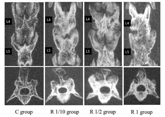
Fig. 1 Computed tomography images of posterolateral lumbar fusion (PLF).
1.CT Examination
Bone fusion rates between the intervertebral joints and transverse processes were analyzed (Fig. 2). At 2 weeks post-surgery, there were no significant differences among the groups. However, from 4 weeks onwards, all three experimental groups (R 1, 1/2, 1/10) showed significantly higher fusion rates compared to the control group (p<0.05). The volume of bone fusion was also assessed (Fig. 3). From 2 weeks post-surgery, all experimental groups demonstrated significantly higher fusion volumes than the control group (p<0.05), although there were no significant differences among the experimental groups themselves. Furthermore, when comparing the rate of volume increase at 2, 4, and 6 weeks postoperatively, all three experimental groups had significantly higher rates than that in the C group (R 1, 1/2, 1/10 group vs. C group; p<0.05) (Fig. 4). In contrast, only the R1 and R1/10 groups had significantly higher rates than that in the C group at 8 weeks postoperatively (R 1, 1/10 group vs. C group; p<0.05).
2.Bone densitometry
The average BMD of the distal femoral diaphysis showed no significant difference among the experimental groups, but all three groups had significantly higher values than that in the C group (R 1, 1/2, 1/10 group vs. C group; p<0.05).
3.Histological examination
Average total PLF areas of the experimental groups at 8 weeks post-surgery showed no significant differences; however, all three groups had significantly larger areas than that of the C group (R 1, 1/2, 1/10 group vs. C group; p<0.05) (Fig. 5). The percentage of bone trabecular area also showed no significant differences among the experimental groups, but all three groups had significantly higher values than that in the C group (R 1, 1/2, 1/10 group vs. C group; p<0.05) (Fig. 6).
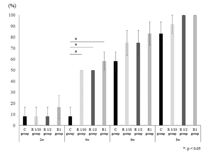
Fig. 2 Bone fusion rates. Bone fusion rates were assessed by micro-CT at 2-week intervals for 8 weeks post-surgery. Data are presented as mean ± SD (n = 6 rats per group) . Statistical analysis was performed using repeated measures ANOVA followed by Tukey’s post-hoc test. Main effect of treatment: F (3, 20) = 15.27, p < 0.001; Main effect of time: F (3, 60) = 268.45, p < 0.001; Interaction: F (9, 60) = 2.18, p = 0.036. *p < 0.05 vs. control group at each time point.
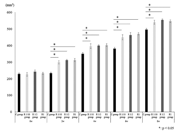
Fig. 3 Posterolateral lumbar fusion volumes. Fusion volumes were measured by micro-CT at 2-week intervals for 8 weeks post-surgery. Data are presented as mean ± SD (n = 6 rats per group) . Statistical analysis was performed using repeated measures ANOVA followed by Tukey’s post-hoc test. Main effect of treatment: F (3, 20) = 178.64, p < 0.001; Main effect of time: F (4, 80) = 2843.73, p < 0.001; Interaction: F (12, 80) = 22.51, p < 0.001. *p < 0.05 vs. control group at each time point.
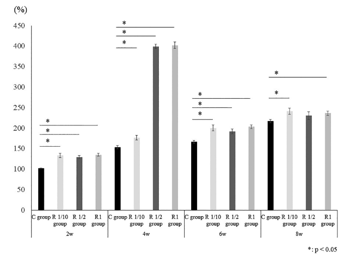
Fig. 4 Rates of posterolateral lumbar fusion volume increases. Rates of fusion volume increases were calculated at 2-week intervals for 8 weeks post-surgery. Data are presented as mean ± SD (n = 6 rats per group) . Statistical analysis was performed using repeated measures ANOVA followed by Tukey’s post-hoc test. Main effect of treatment: F (3, 20) = 1118.68, p < 0.001; Main effect of time: F (3, 60) = 788.95, p < 0.001; Interaction: F (9, 60) = 231.81, p < 0.001. *p < 0.05 vs. control group at each time point.
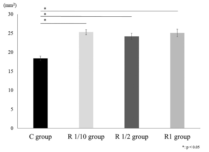
Fig. 5 Posterolateral lumbar fusion (PLF) bone volume. PLF bone volumes were measured at 8 weeks post-surgery. Data are presented as mean ± SD (n = 6 rats per group) . Statistical analysis was performed using one-way ANOVA followed by Tukey’s post-hoc test. F (3, 20) = 139.27, p < 0.001. *p < 0.05 vs. control group.
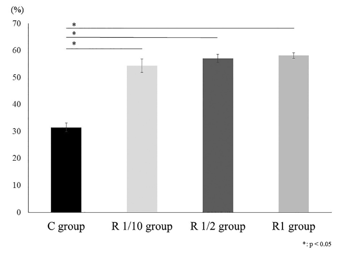
Fig. 6 Trabecular bone area ratios. Trabecular bone area ratios were calculated from histological sections at 8 weeks postsurgery. Data are presented as mean ± SD (n = 6 rats per group). Statistical analysis was performed using one-way ANOVA followed by Tukey’s post-hoc test. F (3, 20) = 351.64, p < 0.001. *p < 0.05 vs. control group.
1.Bone fusion rate
Our analysis of bone fusion rates revealed several key findings:
1.Early effects: No significant differences were observed among treatment groups at 4 weeks post-surgery.
2.Acceleration of bone consolidation: From 4 weeks onward, all experimental groups showed significantly higher osseous integration rates compared to the control group.
3.Dose-independent efficacy: Similar outcomes were observed across all dosages, suggesting that even one-tenth of the previously used dose effectively improved bone fusion rates.
4.Late-stage healing: The lack of significant differences at 6 and 8 weeks post-surgery may be attributed to the inherently high healing rates in rats.
These results suggest that sclerostin antibody administration, regardless of dosage, substantially accelerates bone consolidation, with effects detectable as early as four weeks post-intervention. Kim et al. reported a healing rate of 82.5% for the control group at 8 weeks post-surgery based on CT[9], whereas Kamoda et al. reported a healing rate of 70% for the control group at 8 weeks post-surgery based on radiography[11]. Given this, it is speculated that the high healing rate in the control group and ceiling effect contributed to the lack of significant differences observed.
2.Bone fusion volume
The average volume of bone fusion showed no difference among the groups preoperatively. However, all experimental groups, despite showing no significant differences among them, had significantly larger volumes than that in the C group from 2 weeks onwards. The same trend was observed for the rate of volume increase. These findings suggest that a significant increase in bone volume occurred in all romosozumab-administered groups as early as 2 weeks post-surgery. In addition, similar results observed at different doses imply that even one-tenth of the dosage used in previous studies was effective in increasing the bone fusion volume. Although utilizing a rat fracture model, Ominski et al. reported that the romosozumabadministered group had a 41% larger fracture volume than that in the control group at 7 weeks post-surgery based on micro-CT[12]. Similarly, McDonald et al. reported that the romosozumab-administered group in a rat femoral lengthening model had a 26–38% larger bone regeneration volume than that in the control group at 8 weeks post-surgery[13]. These findings collectively suggest the ability of romosozumab to promote bone proliferation at the graft site.
The primary driver of this phenomenon is believed to be the drug’s effect on skeletal remodeling. The sclerostin antibody binds specifically to its target protein, blocking its interaction with LRP5 and LRP6 receptors. This interference reduces the inhibition of critical cellular signaling pathways. As a result, these signaling pathways become more active in boneforming cells, leading to enhanced osteogenesis and reduced bone breakdown. Previous research has shown that activation of this signaling mechanism is associated with increases in both cortical and trabecular bone mass[5-7]. Therefore, it is speculated that vigorous bone modeling in the graft area contributed to the increased volume and rate of bone fusion observed in all experimental groups compared to the control group.
3.Bone density
Although no significant differences were observed among the experimental groups in terms of the average BMD at the distal femoral diaphysis, all three groups showed significantly higher values than the C group. Numerous reports have documented the effectiveness of romosozumab in increasing bone density[6,8], which was observed in the present study. Furthermore, similar results observed with different doses indicate that even one-tenth of the dosage used in previous studies was effective in increasing bone density.
4.Histological findings
At 8 weeks post-surgery, the average total PLF area showed no significant differences among the experimental groups, but all three groups had significantly larger areas than that in the C group. The percentage of bone trabecular area showed similar results. These histological outcomes can also be attributed to the modeling effect of romosozumab. Trabecular augmentation due to Evenity® has been reported in the clinical data of patients with osteoporosis. Specifically, a sub-analysis of the FRAME trial that assessed bone volume and microarchitecture at 12 months using micro-CT reported a significant increase in trabecular volume fraction with Evenity® administration[6]. Thus, based on the present and previous studies, romosozumab can induce robust remodeling and favorable trabecular formation in bone graft areas of spinal fusion surgeries for patients with osteoporosis. Similar to previous parameters, similar outcomes at different doses implied that even one-tenth of the dosage used in prior studies was effective in producing favorable histological findings.
5.Limitations
Despite the findings of this study, several limitations were noted. One limitation was the lack of bone strength evaluation, such as three-point bending tests, due to the constraints of the mechanical testing equipment. The model used involved only a single intervertebral space, as the amount of grafted bone was insufficient for mechanical testing. In the future, we plan to investigate bone strength using allografts or artificial bones to allow for multiple intervertebral spaces. Another limitation was the evaluation of increased bone density due to romosozumab, which was only performed at the end of the study. This was due to our bone densitometry equipment’s constraints, which require specimen sacrifice and can only measure objects the size of a femur, complicating pre- and post-administration comparisons.
Our study demonstrated the efficacy of romosozumab in promoting bone fusion in a rat PLF model. Romosozumab, though a humanized antibody, has shown cross-reactivity in rats due to the conserved sclerostin sequence[12]. However, we did not directly measure serum sclerostin levels or bone turnover markers. This limitation prevented us from fully elucidating the molecular mechanisms of romosozumab in our model. Future studies should include these measurements to provide more comprehensive evidence of romosozumab’s activity.
Furthermore, it is important to note that the bone fusion-promoting effects of romosozumab in clinical settings remain largely unexplored. Our study provides preliminary evidence in a rat model, but the translation to human patients requires cautious consideration. Future clinical trials should first establish the safety and efficacy of romosozumab in promoting bone fusion in humans, particularly in osteoporotic patients undergoing spinal fusion surgery. These studies should determine the optimal dosage and timing of romosozumab administration in the perioperative period. Additionally, long-term follow-up studies are necessary to assess the durability of fusion and potential adverse effects. Comparative studies with current standard-of-care treatments would also be valuable. As we move towards clinical application, it is crucial to balance the potential benefits of enhanced bone fusion against any risks associated with romosozumab use in this new context.
6.Conclusion
This study investigated the efficacy of lower dosages of romosozumab for promoting bone healing using the same rat lumbar PLF model. Radiographic assessments showed that all antibody-treated groups exhibited significantly improved bone consolidation and increased fusion mass volume compared to the untreated group. Histological examination supported these findings, revealing a substantially higher density of trabecular structures within the fusion region in all antibody-treated groups. Notably, a fractional dose (one-tenth of the standard dose) was as effective as both half- and full-dosage regimens in achieving these positive outcomes.
NT, KI, IT, SuO, YE, YS, and SeO designed research, analyzed, and/or interpreted the data. NT, KI wrote the article and KO, GK, TM, MI, MS-N, TH, TA, SoT, SuT, SO, NS, and SeO gave critical comments on the draft of the manuscript. All authors read and approved the final version of the manuscript.
None.
Seiji Ohtori is one of the Editors of Chiba Medical Journal and on the journal’s Editorial Committee. He was not involved in the editorial evaluation or decision to accept this article for publication at all.
This study was approved by the Ethics Approval Committee of Chiba University School of Medicine (Approval No. A6-039). Informed consent was not required because this study involved no human subject.
Additional data are available via the corresponding author.
Address correspondence to Dr. Kazuhide Inage.
Department of Orthopaedic Surgery, Graduate School of Medicine,
Chiba University, 1-8-1, Inohana, Chuo-ku, Chiba 260-8670, Japan.
Phone: +81-43-226-2117.
Fax: +81-43-226-2116.
E-mail: kazuhideinage@chiba-u.jp
