


Chiba Medical J. 102E:13-17, 2026
doi:10.20776/S03035476-102E-1-P13
〔 Case Report 〕
Junichi Nakamura, Shigeo Hagiwara, Yuya Kawarai
Rui Hirasawa, Jumpei Shoda, and Seiji Ohtori
Department of Orthopedic Surgery, Graduate School of Medicine, Chiba University, Chiba 260-8670.
(Received January 13, 2025, Accepted December 19, 2025, Published March 10, 2026.)
This report presents findings of osseointegration on the metallic surface of a retrieved implant following explantation of a grit blasted and anodized titanium alloy (Ti-6Al-4V) surface on a Zweymüller-type short stem due to deep infection. A 74-year-old woman with a left femoral neck fracture underwent hemiarthroplasty via the direct anterior approach. Despite her history of autoimmune hepatitis, cirrhosis, diabetes, and cancer, she recovered to pre-injury levels of ambulation. However, recurrent cellulitis developed a few months postoperatively. By one year, pain and inability to walk were evident, with imaging showing acetabular bone destruction and prosthesis dislocation. A Girdlestone procedure was performed 1.5 years post-surgery. Prosthesis removal required an extended femoral approach due to strong fixation. Intraoperative cultures identified Streptococcus mitis and Streptococcus lugdunensis. The extracted implant revealed distal bone regeneration. Histopathologic analysis confirmed osseointegration, with bone tissue directly adhering to the implant surface, free of fibrous tissue or inflammation. The findings from the explanted specimen suggest that the 6 - 8 μm surface roughness achieved through grit blasting and anodization supports osseointegration.
Osseointegration, Grit blast, Anodization, Zweymüller stem, Retrieved study
The Zweymüller stem is a cementless stem for total hip arthroplasty designed to achieve initial stability by scratching against the cancellous bone of the femoral canal. It features a grit-blasted surface of titanium alloy (Ti-6Al-4V) with a roughness of 3 μm to 5 μm[1-4]. MIRFY, developed through an industry-academia collaboration among Chiba University, Calm Rana Inc., Surgical Alliance Co., and Mizuho Co, is a purely domestic Japanese hip prosthesis. It inherits the Zweymüller concept while being adapted to the highly curved and shorter femoral shape typical in Japanese women, based on CT data. By shortening the stem length by approximately 2 cm for each size, it aims to facilitate insertion even during minimally invasive surgeries. MIRFY enhances initial stability by employing a grit-blasted surface of Ti-6Al-4V with a roughness increased to 6μm to 8μm. Additionally, it is anodized, resulting in its characteristic dark blue coloration.
Here we report a case involving the explantation of a MIRFY stem due to deep infection and our findings of bone regeneration on the metallic surface of the retrieved implant.
A 74-year-old female presented with a left femoral neck fracture. Pre-injury activities of daily living (ADL) allowed independent ambulation. Her medical history included autoimmune hepatitis with cirrhosis, hyperammonemia, diabetes, post-operative breast cancer, and uterine cervical cancer requiring ureteral stent placement due to adhesions. The initial hemiarthroplasty was performed via a direct anterior approach in the supine position using a mobile traction table (LECURE, Calm Rana Inc., Chiba, Japan and Surgical Alliance Co., Tokyo, Japan) , with the insertion of the MIRFY stem. Postoperatively, her ADL returned to pre-injury levels.
However, several months postoperatively, she began experiencing recurrent episodes of cellulitis. By approximately one-year post-surgery, she developed significant pain and was unable to walk. Radiographic findings revealed acetabular bone destruction and subluxation of the hemiarthroplasty (Fig. 1) . Osteolysis suspicious for periprosthetic infection was identified. The indication for revision surgery following infection remains controversial. In this case, the patient had multiple comorbidities, a compromised general condition, and a decline in ADL to the level of indoor wheelchair use. Three therapeutic options were presented: (1) observation alone, (2) implant removal only (Girdlestone procedure) , and (3) two-stage reconstruction. After a detailed discussion of the advantages and disadvantages of each treatment, the patient elected to undergo Girdlestone procedure 1.5 years postoperatively. The prosthesis removal surgery was performed using the same supine anterior approach as the initial procedure. A radiolucent line was observed proximally around the stem; however, the fixation was strong, necessitating an extended femoral approach with longitudinal femoral splitting for extraction (Fig. 2) . Intraoperative cultures identified Streptococcus mitis and Streptococcus lugdunensis. The infection resolved after intravenous cefazolin 1 g twice daily for 12 days followed by oral levofloxacin 500 mg for two weeks.
Visual inspection of the extracted implant revealed bone regeneration at the distal portion of the stem.
The explanted specimen was embedded in methyl methacrylate resin blocks, and non-decalcified polished sections (40-50 μm thick) were prepared using a specialized polishing system (EXAKT, Germany) . Detached tissue from the implant surface was cut and shaped using ISOMET-1000 (BUEHLER) and sectioned into 6-μm slices using a microtome (LEICA RM2255) . Hematoxylin and Eosin (H&E) , and Villanueva-Goldner-stained sections were prepared. Histopathologic analysis of the adhered regions revealed that the implant-bone interface consisted solely of bone tissue, with no fibrous connective tissue or inflammatory cells interposed, findings consistent with osseointegration (Fig. 3) .

Fig. 1 Imaging progression.
Plain radiographs of the left hip (anteroposterior view) : (a) At the time of the femoral neck fracture; (b) Immediately after hemiarthroplasty; (c) At 1.5 years postoperatively, osteolysis at the lateral acetabular rim led to superior-lateral displacement of the outer cup. Radiolucent lines were noted around the proximal femur, and the hip was malaligned. (d) Corresponding CT image demonstrated bone loss of the posterior acetabular wall with posterior subluxation.

Fig. 2 Intraoperative findings and appearance of the extracted implant. (a)
Intraoperative photograph showing the stem exposed after longitudinal splitting of the femur. Anterior view of the stem (b) , medial view of the distal part (c) , and lateral view of the distal part (d) . The region evaluated histologically is marked by a yellow box.
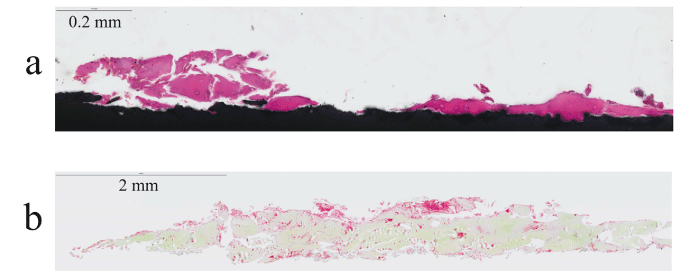
Fig. 3 Histopathologic findings. (a)
H&E staining: Pink areas represent bone tissue. (b) Villanueva Goldner staining: Green areas indicate calcified bone, and red areas indicate osteoid tissue.
The Zweymüller stem is characterized by its rectangular cross-section and surface treatment [1,2]. The sharp edges of the rectangular cross-section achieve fixation by engaging the tubular inner cortex of the femur. The grit-blasted surface promotes bone attachment, described as “bone ongrowth” rather than “bone ingrowth,” as bone tissue overlays the metal surface rather than infiltrating it. The Zweymüller stem demonstrates excellent long-term outcomes. For the Alloclassic stem (Zimmer Biomet, Indiana) , implant survival rates-using revision surgery for any reason as the endpoint-are reported to be 92%-98% 15 years postoperatively. Although data reliability decreases due to a reduced number at risk, survival rates are reported to be 96% at 20 years, 84% at 25 years, and 85% at 30 years[5-8]. The successor model, SL PLUS (Smith & Nephew, Tennessee) also exhibits excellent survival rates of 92%-100% 15 years postoperatively, 85% at 20 years, and 81% at 25 years[9,10]. For the thirdgeneration model, SL PLUS MIA, there is limited long-term data in the English literature. However, a small-scale study involving 41 cases without hydroxyapatite coating reported a 100% survival rate five years postoperatively[11]. However, the clinical outcomes of the hydroxyapatite-coated SL PLUS MIA are yet to be determined[12].
The Zweymüller concept has several advantages. First, ease of use and long-term stability: The design is surgeon-friendly and provides consistent long-term outcomes for both novice and experienced surgeons. Second, wide patient applicability: As a distally fixed stem, it is suitable for elderly osteoporotic patients with weak proximal bone and wide femoral canals. Third, cost efficiency: The simple design reduces production and inventory maintenance costs.
In the research and development of artificial joints, it is crucial to recognize that even minor modifications can pose a risk of poor outcomes. For example, the Elance stem (Kyocera, Kyoto, Japan) , developed as a Zweymüller-type stem, was designed to induce apatite formation on its titanium alloy surface through alkali-heat treatment to promote bone adhesion. However, Tsukada and Wakui reported suboptimal results, with an implant survival rate of only 71.3% five years postoperatively[13]. The grit-blasted surface roughness may have been too fine to achieve sufficient initial fixation strength. With this in mind, the development of the MIRFY stem prioritized differentiation from existing products while adhering to the achievements of predecessors. We decided that our challenge to address was to design a short stem to facilitate easier insertion, even in modern minimally invasive surgeries. The findings from the explanted specimen in this case suggest that the 6-8 μm surface roughness of Ti-6Al-4V achieved through grit blasting and anodization of the MIRFY stem supports osseointegration. This observation holds significant promise for its long-term durability and clinical utility.
Conceptualization: NJ. Funding acquisition: NJ. Investigation: NJ, HS, YK. Methodology: NJ, HR. Supervision: SJ, OS. Visualization NJ, HS. Writing-review & editing: NJ, HS, KY, HR, SJ, OS. All authors read and approved of the final manuscript.
The corresponding author (JN) was supported by JSPS KAKENHI (23K08647) , Hip Joint Foundation of Japan, the Takeda Science Foundation, the Futaba Foundation, Himawari venture Foundation, Chibagin research grant, New Energy and Industrial Technology Development Organization Entrepreneurs Program, Ishikawa Sunrise Industries Creation Organization, Chiba University Venture Business Laboratory research grant, and Health Labor Sciences Research Grant, the Ministry of Health Labor and Welfare, Japan (JPMH23FC0201) .
The corresponding author, NJ, is the founder of Calm Rana Inc. and holds stock in the company. One co-author, SJ, is the Chief Executive Officer Calm Rana Inc. and another co-author, OS is an editorial board member of this journal. The other co-authors have no conflicts of interest to declare.
Written informed consent was obtained from this patient for publication of the present report. This is not human research. This is not animal research.
Not applicable.
Address correspondence to Dr. Junichi Nakamura.
Department of Orthopedic Surgery, Graduate School of
Medicine, Chiba University, 1-8-1, Inohana, Chuo-ku, Chiba
260-8670, Japan.
Phone: +81-43-226-2117.
Fax: +81-43-226-2116.
E-mail: njonedr@chiba-u.jp
