




Chiba Medical J. 102E:63-69, 2026
doi:10.20776/S03035476-102E-3-P63
〔 Case Report 〕
Satoyo Otsuka1) , Hiroshi Ishikawa1) , Haruna Nemoto1) ,
Yusuke Kouchi2) , Ayumu Matsuoka1) , Kyoko Nishikimi1) ,
Shinichi Tate1) , Katsuhiro Nasu3) , Jun-Ichiro Ikeda4) , and Kaori Koga1)
1) Department of Obstetrics and Gynecology, Reproductive Medicine, Graduate School of Medicine, Chiba University, Chiba 260-8670.
2) Department of Molecular Pathology, Graduate School of Medicine, Chiba University, Chiba 260-8670.
3) Comprehensive Radiology Center, Chiba University Hospital, Chiba 260-8677.
4) Department of Diagnostic Pathology, Graduate School of Medicine, Chiba University, Chiba 260-8670.
(Received December 30, 2025, Accepted May 27, 2026, Published September 10, 2026.)
【Purpose】Breast cancer is the most frequent malignant tumor metastasizing to the ovary. Ovarian metastases from breast cancer are commonly bilateral and small, and they are often responsive to anti-estrogen therapy; however, direct invasion of metastatic breast cancer to primary ovarian tumors are extremely rare, and must be differentiated from primary ovarian malignancies.
【Case presentation】An 81-year-old woman with a history of breast cancer surgery 9 years prior was incidentally found to have a 63-mm multilocular tumor in the left ovary. Magnetic resonance imaging revealed that the tumor had diffusion-limiting mural nodules, along with peritoneal dissemination and ascites. While the peritoneal dissemination and ascites responded to anti-estrogen therapy and improved, the ovarian tumor gradually increased in size, and the tumor wall showed thickening with heterogeneous contrast enhancement. Additionally, new cystic lesions were detected in the contralateral ovary. Therefore, we performed a bilateral salpingo-oophorectomy for both diagnostic and therapeutic purposes. Histologically, mammary lobular carcinoma cells extensively infiltrated the cyst wall of the primary ovarian serous cystadenoma, indicating direct invasion of metastatic breast carcinoma into seromucinous cystadenoma in both ovaries.
【Conclusion】Direct invasion of metastatic invasive lobular breast carcinoma to seromucinous cystadenoma in both ovaries exhibited resistance to anti-estrogen therapy and demonstrated progressive enlargement. Multilocular ovarian tumors in patients with a history of breast cancer may reflect direct invasion of metastatic breast cancer into primary ovarian cystic tumors.
breast neoplasms, metachronous neoplasms, mucinous cystadenoma, neoplasm metastasis, ovarian neoplasms
The ovary is one of the potential metastatic sites for breast cancer, following the bone, lung, and liver. The prevalence of metachronous ovarian metastases is 3 - 30% in patients with a history of breast cancer, and the median time from breast cancer onset to ovarian metastasis is approximately 5 years [1]. Ovarian metastasis from breast cancer typically presents as bilateral and small (< 5 cm) lesions, with predominantly enhancing components and occasional cysts, and the most common histological type is invasive lobular carcinoma (ILC) [2]. Ovarian metastases from breast cancer are often asymptomatic and incidentally detected. Breast cancer typically metastasizes to the ovarian stroma; however, direct invasion into an ovarian tumor is extremely rare.
Anti-estrogen therapy is generally performed for the treatment of recurrence or metachronous metastasis of hormone-responsive breast cancer. ILC is highly positive for estrogen receptor (ER) and progesterone receptor (PgR) and is responsive to anti-estrogen therapy[3]. Therefore, ovarian tumors with direct invasion by metastatic ILC may shrink in response to anti-estrogen therapy. Herein, we report the case of direct invasion of metastatic breast carcinoma into primary seromucinous cystadenoma of both ovaries that enlarged following anti-estrogen therapy, in a patient with a history of breast cancer.
An 81-year-old woman was referred to our department for further examination due to an elevated serum cancer antigen 125 (CA125) level and an incidentally detected ovarian tumor. The patient underwent a right mastectomy and sentinel lymph node biopsy for locally advanced breast cancer 9 years earlier and was diagnosed with ILC (Supplemental Fig. 1a) . The postoperative TNM classification was pT3N0M0. Neoplastic cells were positive for ER and PgR, and the membranous expression of E-cadherin was reduced and became indistinct (Supplemental Fig. 1b) . Sentinel lymph node metastasis was negative. The patient was treated with letrozole postoperatively, and then tamoxifen citrate was administered 12 months later for a total duration of 60 months. The patient’s general conditions remained stable, without any signs of recurrence. The patient underwent pelvic magnetic resonance imaging (MRI) 83 months after breast cancer surgery to assess a contusion from a fall, and a 63-mm tumor was incidentally identified in the left ovary. At this time, the mass was presumed to be a benign ovarian cystic tumor.
Seven months later, a contrast-enhanced MRI revealed that the multilocular tumor had increased in size to 71 mm, with newly detected ascites and peritoneal thickening, indicating possible intraabdominal cancer dissemination; however, the tumor did not show any solid components with enhancement (Fig. 1a) . The serum CA125 level gradually increased, and ascitic fluid cytology of the cell block revealed the atypical cells to be immunoreactive for ER and GATAbinding protein 3 (GATA3) , a specific antibody for breast cancer, and negative for human epidermal growth receptor type 2 (HER2) , which may be expressed in breast cancer, suggesting a recurrence of breast cancer (Supplemental Fig. 2) . Therefore, she was administered fulvestrant as an anti-estrogen agent for 11 months. The peritoneal thickening, which was indicative of peritoneal dissemination, shrank in parallel with the decrease in the proportion of ascites. On the other hand, the size of the left ovarian tumor further increased up to 127 mm, and MRI revealed small nodules with diffusion restriction and contrast enhancement in the tumor (Fig. 1b, 1c, 1d) . Additionally, the right ovary, which had shown clustered small cysts but was not recognized as pathological on the initial MRI, had also obviously increased in size with cystic lesions (Fig. 1e, 1f) . Based on the discrepancy in the response to the anti-estrogen therapy between the peritoneal lesions and ovarian tumors, we suspected primary ovarian borderline tumors rather than metastatic ovarian tumors and performed bilateral salpingo-oophorectomy and partial omentectomy.
No apparent peritoneal dissemination was observed at laparotomy. The left ovary was occupied by an oligocystic lesion measuring 14.0 cm in a major diameter (Fig. 2a) . The right ovary measured 3.2 cm × 3.0 cm × 1.4 cm and contained small cysts up to 0.5 cm in length. The cystic lesions in both ovaries had mucopurulent content, irregularly thickened cyst walls, and numerous whitish, firm, and plaque-like lesions on the luminal and external surfaces (Fig. 2b, 2c) . The omentum was slightly thickened with white fibrosis (Fig. 2d) . Histologically, ILC metastases were observed in the bilateral ovaries and fallopian tubes, peritoneum, and omentum. Moreover, a primary seromucinous cystadenoma was detected in both ovaries. ILC metastases were directly infiltrating the cyst wall of the primary ovarian serous cystadenoma, indicating direct invasion of metastatic breast carcinoma into seromucinous cystadenoma (Fig. 3a) . In the areas of the metastatic ILC, small monotonous neoplastic cells with pale eosinophilic cytoplasm infiltration, showing various architectural patterns including macronodular, diffuse/solid, single file, and small nests/ clusters were observed (Fig. 3b-3d) . The neoplastic cells were positive for ER, GATA3, and gross cystic disease fluid protein 15 (GCDFP-15) , while almost negative for E-cadherin and Paired Box 8 (PAX8) (Fig. 3e, 3f) , consistent with those of ovarian metastasis from mammary ILC. In contrast, the lining cells of the lumens were composed of endocervical-like mucinous, serous, and eosinophilic indifferent cells in the seromucinous cystadenoma components of both ovaries (Fig. 3g) . Cytological atypia of the lining cells was absent, and neither borderline nor malignant component was identified. Therefore, we diagnosed direct invasion of metastatic breast carcinoma into seromucinous cystadenoma in both ovaries. Although the patient continued to receive fulvestrant after bilateral salpingo- oophorectomy, peritoneal dissemination worsened. The patient was then administered palbociclib but died of breast cancer 104 months after the initial surgery.
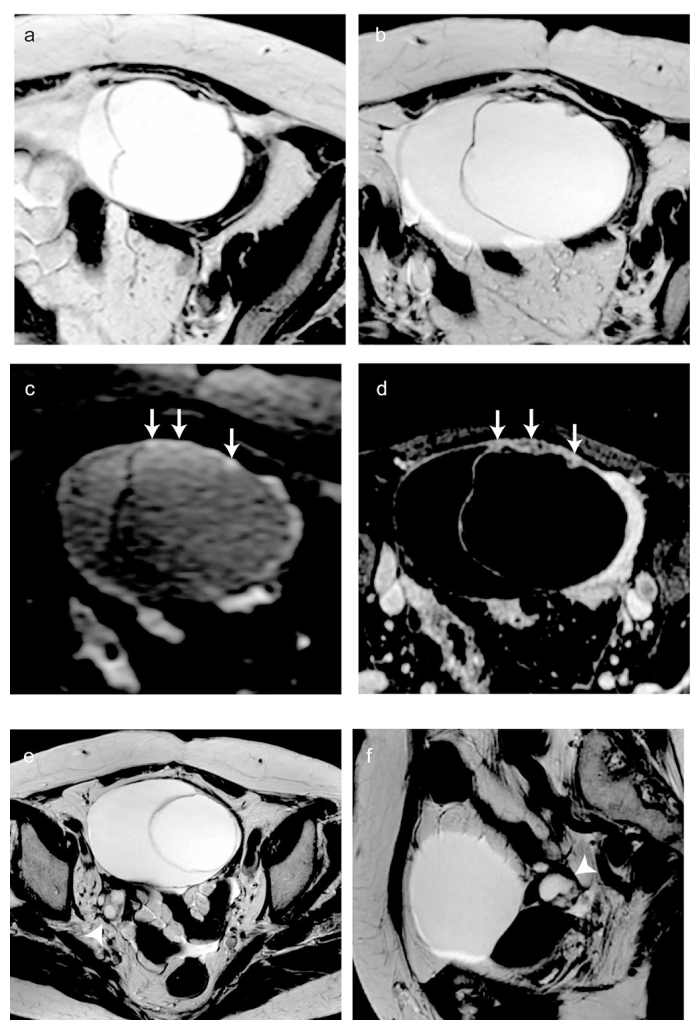
Fig. 1 Magnetic resonance imaging (MRI) findings of the ovarian tumor. (a-d) : MR images of the left ovarian tumor. (a) T2-weighted axial image at the initial study. The left ovarian tumor is a multilocular cystic mass measuring 71 mm in maximum diameter, with thin walls and septa. (b) T2-weighted axial image eight months after the initial study. The left ovarian tumor has increased to 127 mm in maximum diameter, and wall thickening has appeared on the ventral aspect. Multiple microcysts are confirmed within the thickened wall (black arrows) . (c) Diffusion weighted image. The solid portion of the thickened wall shows high signal intensity (white arrows) . (d) Postcontrast fat-suppressed T1-weighted image. The solid portion shows heterogeneous contrast enhancement (white arrowheads) . (e-f) MR images of the right ovary. (e) T2-weighted axial image at the initial study. The right ovary measured 21 mm in maximum diameter with multiple small cysts (black arrowhead) . Although the size and characteristics were incongruous for a woman in her eighties, however, this finding was not recognized as pathological at the time. (f) T2-weighted axial image 8 months after the initial study. The right ovary has not shown obvious enlargement (black arrowhead) ; however, the internal small cysts are confirmed to have increased in size.
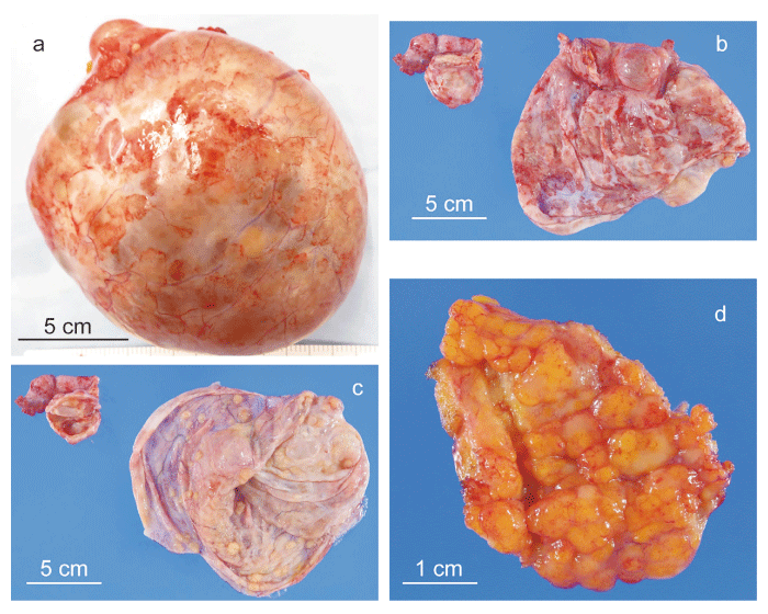
Fig. 2 Gross appearance of the resected specimens. (a) Resected left ovarian tumor. The surface is smooth, and no adhesions to the surrounding tissues are observed. The maximum diameter of the tumor had increased to 14 cm. (b) (c) Cut surfaces of the resected bilateral ovarian tumors. Macroscopically, the bilateral ovarian tumors appear as cystic lesions with irregularly thickened walls and numerous nodules and plaque-like lesions on the internal and external surfaces. (d) Resected thickened omentum. Part of the resected omentum is thickened, with a palpable, firm nodule.
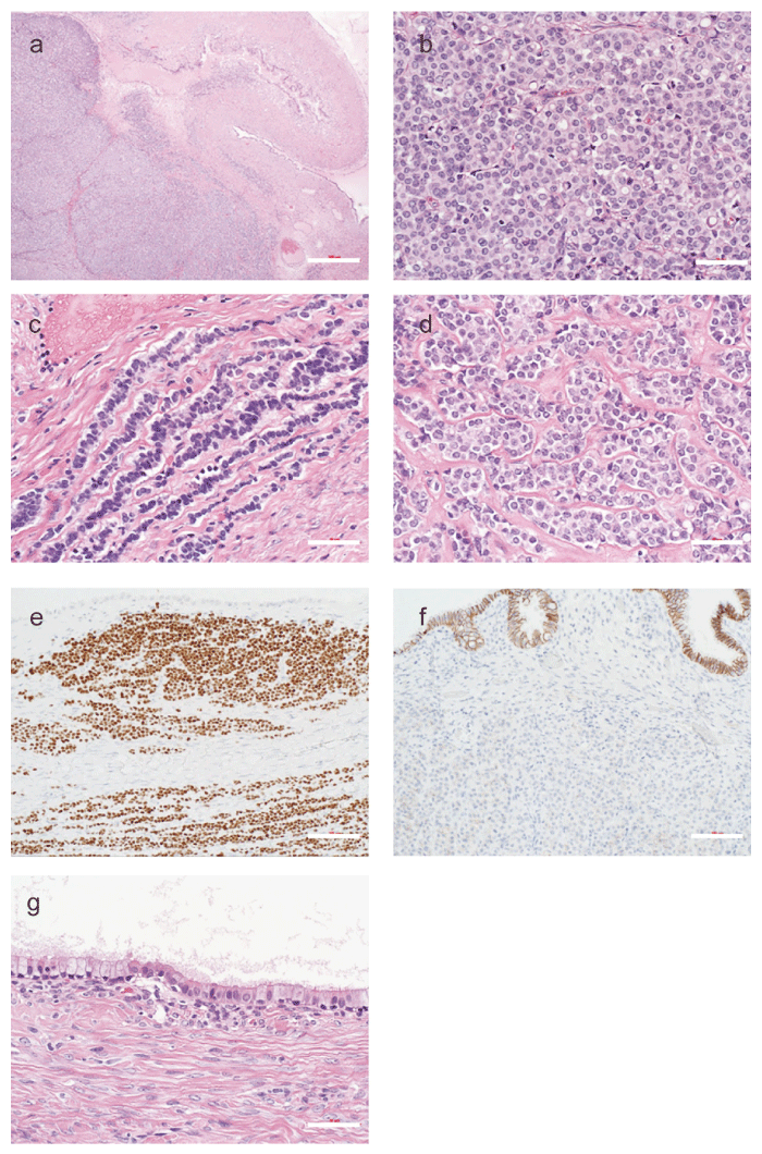
Fig. 3 Histological and immunohistochemical analysis of the left ovarian tumor. (a) Low-power view of Hematoxylin and eosin (HE) staining of the left ovarian tumor. Scale bar = 500 μm. Bilateral ovarian tumors reveal collision tumors of metastatic invasive lobular carcinoma (ILC) of the breast and seromucinous cystadenoma. The upper right area shows seromucinous cystadenoma, whereas the lower left area shows metastatic ILC. (b) (c) (d) High-power view of HE staining of the left ovarian tumor. Scale bar = 50 μm. In the metastatic ILC components, the neoplastic cells infiltrated diffuse/solid (b) , single-file (c) , and small nested/clustered pattern (d) . (e) Immunohistochemistry for GATA-binding protein 3 (GATA-3) of the left ovarian tumor. Scale bar = 100 μm. Immunostaining for GATA3 was diffusely positive. These neoplastic cells are entirely small, monotonous, and pale eosinophilic. Intracytoplasmic lumina are occasionally observed. (f) Immunohistochemistry for E-cadherin of the left ovarian tumor. Scale bar = 100 μm. The luminal epithelial cells of the cystadenoma component show positive membranous staining, serving as an internal control, whereas the metastatic ILC cells in the stroma are negative for E-cadherin, supporting the diagnosis of metastatic breast ILC. (g) High-power view of HE staining of the left ovarian tumor. Scale bar = 50 μm. In the seromucinous cystadenoma components, the lining cells of the cyst walls are composed of endocervical-like mucinous cells and serous (ciliated) cells.
Breast cancer is one of the most frequent tumors to metastasize to ovaries. Although mammary ILCs are associated with endocrine organ metastasis, including the ovary and peritoneal dissemination, coexistence of mammary ILC metastasis with primary benign ovarian tumors is a rare condition. To the best of our knowledge, direct invasion of a metastatic mammary ILC into benign ovarian cystadenomas has not been reported. Previous reports featuring the coexistence of metastasis of mammary ILC with benign ovarian tumors have revealed that histological types of benign ovarian tumors are diverse, such as teratoma and Brenner tumor, and the reasons for detecting the ovarian tumor varied, ranging from systemic evaluations for breast cancer recurrence to incidental findings on imaging performed for unrelated indications [4-6]. The reported gross findings of the lesion are often similar to those of a coexisting benign tumor. In the present case, the left ovarian tumor appeared as a large oligocystic mass, consistent with as previously reported seromucinous cystadenomas [7].
Patients with a history of breast cancer are at increased risk of developing primary malignant ovarian tumors[2]. In addition, hereditary breast and ovarian cancer syndrome associated with BRCA mutations is known to increase the risk of ovarian cancer; however, BRCA genetic testing was not performed in this patient. Since treatment strategies are entirely different between metastatic breast cancer and primary ovarian malignancies, both differentiations are crucial in patients with a history of breast cancer. In this case, a differential diagnosis between ovarian metastasis of breast cancer and ovarian serous borderline tumor was required. One of the key differentiating points between ovarian metastases from breast cancer and primary borderline ovarian tumors lies in their imaging characteristics. Borderline ovarian tumors typically present as cystic lesions or as cystic masses with well-demarcated solid components, and are usually unilateral [8]. In contrast, ovarian metastases from breast cancer are predominantly solid and bilateral [2]. In the current case, preoperative imaging predominantly highlighted the rapidly enlarging left ovarian tumor, and the relatively enlarged, albeit small, right ovarian tumor for the patient’s age was overlooked. In addition, a discrepancy was observed in the effect of anti-estrogen therapy between the peritoneal dissemination and the left ovarian tumor. While the peritoneal dissemination improved and the proportion of ascites reduced, the size of the left ovarian lesion increased. Anti-estrogen therapy was effective for peritoneal dissemination but not for ovarian metastases. Moreover, direct invasion of metastatic mammary ILC into seromucinous cystadenoma is extremely rare. These findings led to the suspicion of a primary borderline tumor rather than ovarian metastasis from breast cancer prior to surgery. In this case, the patient’s performance status was good, and surgery was performed to establish a definitive diagnosis. In similar situations, observation may also be an option for patients with poor performance status.
Immunohistochemical analysis of ovarian tumors is essential in distinguishing between ovarian metastases of breast cancer and primary ovarian tumors. The distinction between the two can be facilitated using a specific combination of immunohistochemical markers [2]. Positive immunoreactivity for GATA3, GCDFP-15, and mammaglobin is characteristic of breast cancerderived tumors, whereas positivity for Wilms’ tumor antigen-1 (WT1) , CA-125, and PAX8 suggests a primary ovarian origin. The detection of ovarian metastases using a combination of immunohistochemical markers has also been reported[9]. Combining the targets E-cadherin, epithelial membrane antigen (EMA) , and Her2/neu resulted in nearly 100% detection of ductal ovarian metastases, and the combination of EMA, Her2/neu, and epithelial cell adhesion molecule was most suitable for detecting lobular ovarian metastases. In the current case, the neoplastic cells were positive for ER, GATA3, and GCDFP-15, while almost negative for E-cadherin and PAX8, consistent with those of ovarian metastasis from mammary ILC.
In summary, we encountered a case of metachronous direct invasion of metastatic breast carcinoma directly into ovarian seromucinous cystadenoma in both ovaries. In contrast to the response to anti-estrogen therapy for peritoneal dissemination and ascites, the cystic portion continued to enlarge, and small nodules demonstrating contrast enhancement emerged within the cyst wall. Direct invasion of metastatic breast cancer into the ovarian tumor is extremely rare, and characteristic MRI and histopathological findings contributed to the diagnosis.
All authors contributed to the study conception and design. Material preparation, data collection and analysis were performed by [S.O.], [H.N.], [Y.K.] [K.N.] and [H.I.]. The first draft of the manuscript was written by [S.O.], [H.N.] and [Y.K.]. The manuscript was reviewed and edited by [H.I.]. Funding acquisition was performed by [K.K.]. Supervision of the manuscript was performed by [S.T.] and [K.K.]. All authors commented on previous versions of the manuscript. All authors read and approved the final manuscript.
This research received no specific grant from any funding agency in the public, commercial, or not-forprofit sectors.
The authors declared no conflict of interest.
Written informed consent was obtained from the patient’s family for publication of this case report and accompanying images. A copy of the written consent is available for review by the Editor-in-Chief of this journal on request.
The data underlying this article are available from the corresponding author upon reasonable request, subject to patient privacy and institutional regulations.
We would like to thank Editage (www.editage.com) for English language editing. We thank Dr. Takeshi Nagashima, Department of Breast Surgery, Chiba University Hospital for the treatment for breast cancer, Dr. Akiyo Takada for diagnostic imaging and the patient’s family for allowing the publication of the manuscript and any accompanying images.
Address correspondence to Dr. Satoyo Otsuka.
Department of Obstetrics and Gynecology, Reproductive Medicine,
Graduate School of Medicine, Chiba University, 1-8-1 Inohana, Chuou-Ku, Chiba 260-8670, Japan.
Phone: +81-43-226-2121.
Fax: +81-43-226-2122.
E-mail: satoyo.1121@gmail.com
