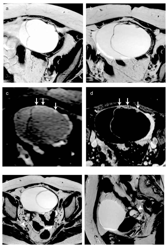
Fig. 1 Magnetic resonance imaging (MRI) findings of the ovarian tumor. (a-d) : MR images of the left ovarian tumor. (a) T2-weighted axial image at the initial study. The left ovarian tumor is a multilocular cystic mass measuring 71 mm in maximum diameter, with thin walls and septa. (b) T2-weighted axial image eight months after the initial study. The left ovarian tumor has increased to 127 mm in maximum diameter, and wall thickening has appeared on the ventral aspect. Multiple microcysts are confirmed within the thickened wall (black arrows) . (c) Diffusion weighted image. The solid portion of the thickened wall shows high signal intensity (white arrows) . (d) Postcontrast fat-suppressed T1-weighted image. The solid portion shows heterogeneous contrast enhancement (white arrowheads) . (e-f) MR images of the right ovary. (e) T2-weighted axial image at the initial study. The right ovary measured 21 mm in maximum diameter with multiple small cysts (black arrowhead) . Although the size and characteristics were incongruous for a woman in her eighties, however, this finding was not recognized as pathological at the time. (f) T2-weighted axial image 8 months after the initial study. The right ovary has not shown obvious enlargement (black arrowhead) ; however, the internal small cysts are confirmed to have increased in size.