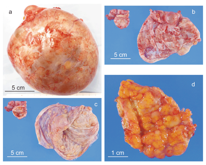
Fig. 2 Gross appearance of the resected specimens. (a) Resected left ovarian tumor. The surface is smooth, and no adhesions to the surrounding tissues are observed. The maximum diameter of the tumor had increased to 14 cm. (b) (c) Cut surfaces of the resected bilateral ovarian tumors. Macroscopically, the bilateral ovarian tumors appear as cystic lesions with irregularly thickened walls and numerous nodules and plaque-like lesions on the internal and external surfaces. (d) Resected thickened omentum. Part of the resected omentum is thickened, with a palpable, firm nodule.