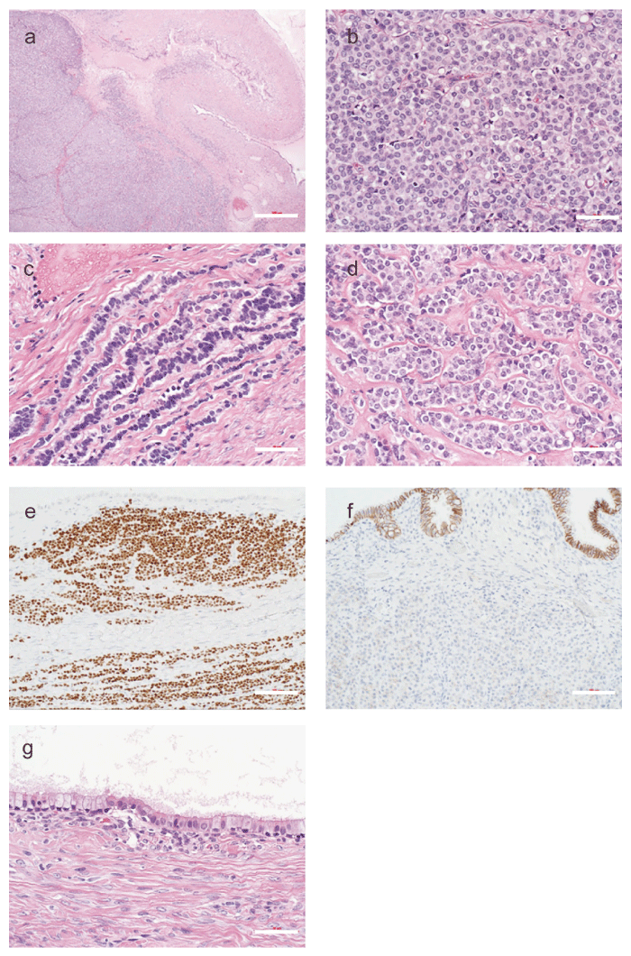
Fig. 3 Histological and immunohistochemical analysis of the left ovarian tumor. (a) Low-power view of Hematoxylin and eosin (HE) staining of the left ovarian tumor. Scale bar = 500 μm. Bilateral ovarian tumors reveal collision tumors of metastatic invasive lobular carcinoma (ILC) of the breast and seromucinous cystadenoma. The upper right area shows seromucinous cystadenoma, whereas the lower left area shows metastatic ILC. (b) (c) (d) High-power view of HE staining of the left ovarian tumor. Scale bar = 50 μm. In the metastatic ILC components, the neoplastic cells infiltrated diffuse/solid (b) , single-file (c) , and small nested/clustered pattern (d) . (e) Immunohistochemistry for GATA-binding protein 3 (GATA-3) of the left ovarian tumor. Scale bar = 100 μm. Immunostaining for GATA3 was diffusely positive. These neoplastic cells are entirely small, monotonous, and pale eosinophilic. Intracytoplasmic lumina are occasionally observed. (f) Immunohistochemistry for E-cadherin of the left ovarian tumor. Scale bar = 100 μm. The luminal epithelial cells of the cystadenoma component show positive membranous staining, serving as an internal control, whereas the metastatic ILC cells in the stroma are negative for E-cadherin, supporting the diagnosis of metastatic breast ILC. (g) High-power view of HE staining of the left ovarian tumor. Scale bar = 50 μm. In the seromucinous cystadenoma components, the lining cells of the cyst walls are composed of endocervical-like mucinous cells and serous (ciliated) cells.