


Chiba Medical J. 93E:11~15,2017
doi:10.20776/S03035476-93E-1-P11
[ Case Report ]
Keisuke Koroki1), Akitoshi Kobayashi1), Akinari, Miyazaki2)
Atsuyoshi Seki1),Seiko Togo1), Takeshi Ando1),
Takashi Maruyama2), and Hideaki Mizumoto1)
1) Department of Gastroenterology, Funabashi Municipal Medical Center, Funabashi 273-8588
2) Department of Surgery, Funabashi Municipal Medical Center, Funabashi 273-8588
(Received June 15, 2016, Accepted July 19, 2016)
We report the case of a successful percutaneus endoscopic necrosectomy for extensive infected walled-off necrosis(WON) followed by acute pancreatitis. A 69-year-old man was examined at our hospital for the chief complaint of upper abdominal pain for the past several weeks. Abdominal computed tomography scanning showed necrotizing pancreatitis with a gas pattern and an extensive necrosis extending to the pelvic cavity. A percutaneous drainage regimen was adopted because it was determined that the amount of fluid accumulation and the area covered by the necrotic lesion were too extensive to cope with drainage through a transgastric route alone using an endoscopic ultrasonography. However, because the lesion had already developed infected WON, we determined that the infection could not be controlled unless the necrotic tissue was removed. Hence, an endoscopic necrosectomy using grasping forceps and retrieval net was performed. Upon completion of the necrosectomy, a decrease in the amount of necrotic tissue was observed through both endoscopy and abdominal computed tomography. While endoscopic necrosectomy and the step-up approach tend to be recommended, percutaneous treatment or conventional open necrosectomy may be required when the range of necrosis is extensive, as in our case. Thus, it appears important to respond flexibly to each case.
walled-off necrosis, necrotizing pancreatitis, endoscopic necrosectomy
With regard to local complications followed by acute pancreatitis, the revised Atlanta classification refers to the encapsulated liquefaction of necrotic pancreatic or peripancreatic tissue four weeks after the onset of acute pancreatitis as“ walled-off necrosis”(WON)[1]. For the treatment of infected WON in particular, drainage alone is insufficient and necrosectomy is required[2]. In recent years, excellent treatment outcomes have been reported with minimally invasive treatments, such as endoscopic necrosectomy, in place of conventional open necrosectomy[3-7]. Herein, we report the case of a successful percutaneus endoscopic necrosectomy performed for extensive infected WON followed by acute pancreatitis.
A 69-year-old man was examined at our hospital for the complaint of upper abdominal pain for the past several weeks. He had no significant past medical or surgical history. He ingested alcohol daily(approximately 80 g/day). The patient's body temperature was 37.6℃, blood pressure was 103/68 mmHg, pulse rate was regular at 117 bpm, SpO2 was 96%(room air), and he was conscious and alert. On physical examination, he had mild upper abdomen tenderness. Laboratory findings on admission showed that an inflammatory reaction was observed with a white blood cell count of 29100/μL and a C-reactive protein level of 18.4 mg/dL. Abdominal computed tomography showed necrotizing pancreatitis with a gas pattern and an extensive necrosis extending to the pelvic cavity(Fig. 1).
Table. 1
Laboratory data on admission
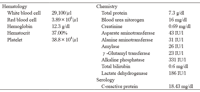
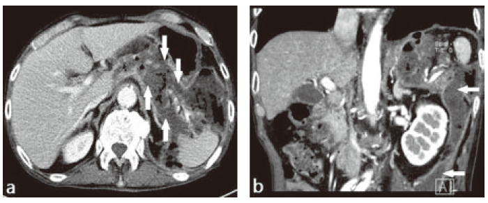 Fig. 1 Abdominal CT scan. Necrotizing pancreatitis with a gas pattern and an extensive necrosis extending to the pelvic cavity.
Consequently, the patient was diagnosed with infectious pancreatic necrosis, and treatment was initiated with fasting and antibiotics. A percutaneous drainage was adopted because it was determined that the amount of fluid accumulation and the area covered by the necrotic lesion were too extensive to cope with drainage through a transgastric route alone using an endoscopic ultrasonography. Initially, two 14-Fr drainage tubes were placed, after which they were replaced by tubes with progressively larger diameters increasing to 18- and then 22-Fr(Fig. 2). However, because the lesion had already developed infected WON, we determined that the infection could not be controlled unless the necrotic tissue was removed. Hence, a necrosectomy was performed.
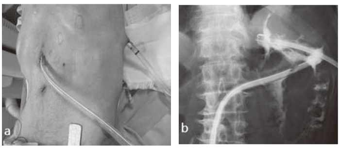 Fig. 2 Initially, two 14-Fr drainage tubes were placed, after which they were replaced by tubes with progressively larger diameters increasing to 18- and then 22-Fr.
On the 24day of hospitalization, the first necrosectomy was performed. An endoscope(GIF-XP260N, Olympus, Japan, diameter: 5.5 mm) was percutaneously inserted into the WON, and necrotic tissue was removed using biopsy forceps(Radial Jaw 4 Pediatric with needle,Boston Scientific, Japan) for endoscopy.
With this set-up, we successfully removed a small amount of the necrotic tissue. After we began using 28- Fr diameter drainage tubes, we were able to insert an endoscope(GIF-Q260, Olympus, Japan, diameter: 9.2 mm). We performed the second and third necrosectomy on the 31 and 38 day of hospitalization, respectively. After inserting this endoscope into the WON, we successfully removed a large amount of necrotic tissue using grasping forceps (FG-8L-1 and FG-45L-1, Olympus, Japan), a retrieval net(Roth Net -foreign body- standard, Olympus, Japan), and other instruments (Fig. 3).Upon completion of the third necrosectomy, a decrease in the amount of necrotic tissue was observed through both endoscopy and abdominal computed tomography(Fig. 4).
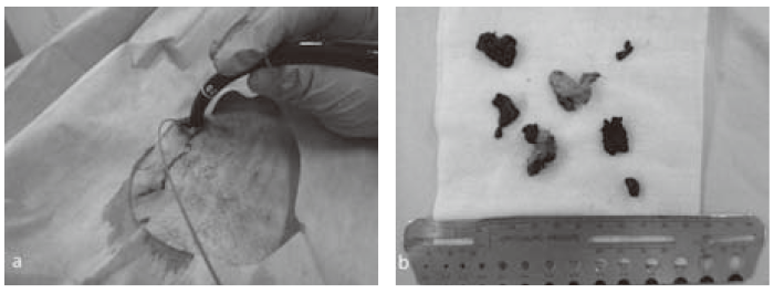 Fig. 3a After we began using 28-Fr diameter drainage tubes, we were able to insert a standard peroral endoscope (GIF-Q260, Olympus, Japan, diameter: 9.2 mm).
Fig. 3b We successfully removed a large portion of necrotic tissue using grasping forceps(FG-8L-1 and FG-45L-1, Olympus, Japan), a retrieval net (Roth Net -foreign body- standard, Olympus, Japan), and other instruments.
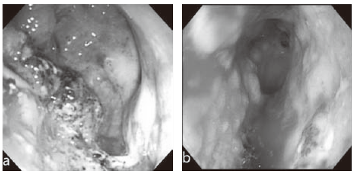 Fig. 4a Before the necrosectomy.
Fig. 4b After the third necrosectomy. A decrease in the amount of necrotic tissue was observed through endoscopy.
When the Atlanta classification was revised in 2012, fluid accumulation, a local complication of acute pancreatitis, was classified into acute peripancreatic fluid collection, acute necrotic collection (ANC), pancreatic pseudocyst, and WON, depending on the presence of necrosis and the time-course since onset[1]. ANC usually develops into WON from four weeks of onset. At the time of admission, we thought our patient was in the state of ANC, the deposits of which became subsequently encapsulated and resulted in WON.
Various procedures for treatment of WON have been reported, including surgical, percutaneous, and endoscopic procedures. Of these, the effectiveness of minimally invasive endoscopic treatment in particular has been widely reported in recent years. Van Sanvoort et al. reported that approximately 60% of WON cases can be treated with drainage alone[8]; however, in cases where drainage alone does not lead to successful treatment, necrosectomy should be considered. In our patient, less invasive percutaneous drainage was first performed after admission.
Garner et al. retrospectively compared treatment outcomes of both endoscopic transgastrointestinal drainage and endoscopic necrosectomy for WON; they reported that in those requiring necrosectomy, a higher rate of subjects were associated with infection[2]. Their results showed that in WON associated with infection, as in our patient, treatment based on drainage alone is often insufficient and necrosectomy is required. Likewise, we performed necrosectomy following drainage, as well. Results of a randomized controlled study have been reported regarding the“ step-up approach,” in which necrosectomy is performed after drainage as necessary[3]. According to this report, percutaneous drainage was performed first in the step-up approach group, and when no improvement was obtained, minimally invasive retroperitoneal debridement was performed. Lower rates of complications and mortality were reported in subjects percutaneously treated using the step-up approach versus those treated using conventional open necrosectomy. Thus, we continued treatment of our patient by avoiding open necrosectomy, whenever possible.
With regard to necrosectomy procedures, the effectiveness of endoscopic necrosectomy has been recently reported. In a randomized controlled trial, rates of serious accidental symptoms and mortality were significantly lower with endoscopic than with conventional open necrosectomy[4]. Furthermore, there have been multiple reports on endoscopic necrosectomy performed for infectious WON, with success rates of 80% and 75% based on reports from Germany[5] and Japan[6], respectively.
In the present study, we initially used an endoscope with a small diameter(5.5 mm) for necrosectomy, which limited usable devices to those that could be inserted through the forceps channel and resulted in insufficient removal of necrotic tissues. Replacement with an endoscope of a standard diameter(9.2 mm) enabled us to remove a large quantity of necrotic tissue. Based on these findings, we concluded that the choice of endoscope is crucial in necrosectomy.
Although there are many reports highlighting the effectiveness of endoscopic necrosectomy[13-18], in cases where the lesion has extended to the pelvic cavity or its peripheral regions, only transgastrointestinal treatment is likely insufficient and a percutaneous approach may be required. The usefulness of a hybrid approach in which percutaneous treatment is used in combination with transgastrointestinal treatment has been reported[9,19]. Furthermore, the effectiveness of the treatment in which endoscopic necrosectomy is performed through a percutaneous approach has also been reported[20].
Because the extent and range of WON in our patient was such that it would definitively require a percutaneous approach later, we decided to perform percutaneous treatment from the beginning itself. Access routes for percutaneous drainage and necrosectomy include transabdominal and retroperitoneal routes. Procedures and routes are chosen at the discretion of the operator, depending on the site of WON. The retroperitoneal approach has been shown to be associated with fewer complications than the open necrosectomy[10,11]. However, it remains unclear whether the transgastrointestinal step-up approach or percutaneous drainage with subsequent percutaneous necrosectomy is more effective. Currently, a relevant randomized controlled trial is being conducted[12]. In the current patient, percutaneous necrosectomy was safely performed, and infected WON was successfully controlled. While endoscopic necrosectomy and the step-up approach tend to be recommended, percutaneous treatment or conventional open necrosectomy may be required when the range of necrosis is extensive, as in our case. Thus, it appears important to respond flexibly to each case.
Address correspondence to Dr. Keisuke Koroki.
Department of Internal Medicine, Funabashi Municipal Medical Center, 1-21-1, Kanasugi, Funabashi-shi, Chiba 273-8588, Japan.
Phone: +81-47-438-3321. Fax: +81-47-438-7323.
E-mail: k.k.cricket.7@gmail.com
