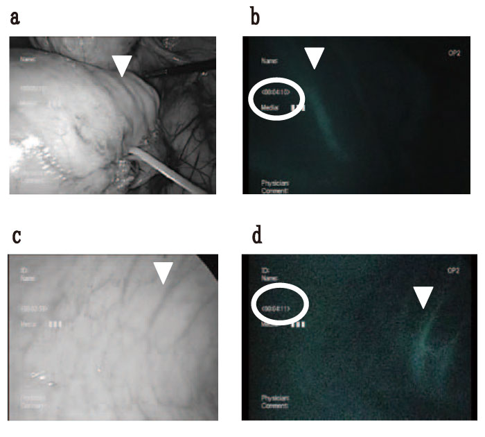
Fig. 1 An NIR perfusion assessment in a Pig undergoing laparoscopic low anterior resection.
We compared the two approaches simultaneously (the same time is shown in the red circle) in the pig experiments. The arrow indicates the same artery of the marginal colon. a/b: Laparoscopic view. c/d: Transanal view.
a. The laparoscopic view under normal light. The SRA is taped.
b. The laparoscopic view under near-infrared fluorescence. Good blood flow of the marginal artery can be confirmed.
c. The transanal view under white light.
d. The trans-anal view under near-infrared fluorescence. Good flow of the capillaries can be confirmed.
b/d. It was compared to the same vessel with the arrow from both approaches using the Image J software program. The gray value of the transanal approach is 77 and abdominal approach is 45.