


Chiba Medical J. 95E:45-52,2019
doi:10.20776/S03035476-95E-3-P45
[ Original Article ]
Junichi Nakamura1), Koh Shimizu2), Toru Suguro3)
Shigeo Hagiwara1), Sumihisa Orita1), Tsutomu Akazawa4),
Takayuki Nakajima5), Yawara Eguchi6), Kazuhide Inage1),
Yasuhiro Shiga1),and Seiji Ohtori1)
1)Department of Orthopaedic Surgery, Graduate school of Medicine, Chiba University, Chiba 260-8670 .
2)Department of Orthopaedic Surgery, Chiba rosai Hospital, Chiba 290-0003 .
3)Japanese Artificial joint Institute, Chiba 292-0036 .
4)Department of Orthopaedic Surgery, St. Marianna University School of Medicine, Kanagawa 216-8511 .
5)Department of Orthopaedic Surgery, Eastern Chiba Medical Center, Chiba 283-8686 .
6)Department of Orthopaedic Surgery, Shimoshizu National Hospital, Chiba 284-0003 .
(Received December 29, 2018, Accepted February 8, 2019, Published June 10, 2019.)
The aim of this prospective cohort study was to document the preliminary results of the direct medial approach, a novel surgical technique for primary total knee arthroplasty. From September 2015 to May 2018, 100 patients were consecutively registered. The inclusion criteria were a primary total knee arthroplasty via the direct medial approach performed by the first author(consultant group)or by residents supervised by the first author(residents group). The follow-up period was three months. The essence of the surgical technique was a medial oblique skin incision of 14 cm from the medial aspect of the tibial tuberosity along the mid-point of the muscle belly of the vastus medialis. The deep synovial layer was preserved in a V-shaped flap, which was useful to reconstruct the medial capsule. Ten percent of the patients had severe adverse events, but all complications completely resolved conservatively and no revision surgeries were required. The complication rates were similar for the consultant and the residents. Surgical time was significantly shorter for the consultant than the residents(92±13[ mean±standard deviation] minutes versus 104±15 minutes, p=0.001). Postoperative range of motion was significantly larger in patients operated on by the consultant than by the residents(124±13 degrees versus 120±11 degrees, p=0.026). Less postoperative pain, larger preoperative range of motion, and surgery performed by the consultant were predictive factors for better postoperative range of motion. This preliminary study demonstrated that the direct medial approach would be safe and effective for primary total knee arthroplasty.
direct medial approach, primary total knee arthroplasty, preliminary results, a novel surgical technique, a prospective cohort study
The subvastus(Southern)approach for total knee arthroplasty(TKA)was first described by Hofmann et al.[1]in 1991. This anatomical technique eliminates disassociation and dehiscence of the vastus medialis obliquus muscle(VMO). The patellar tracking is natural because this approach preserves the integrity of the extensor mechanism and the vascular supply to the patella. The subvastus approach has several advantages, but it has not been popular in clinical practice due to its technical difficulty. Recently, this approach has attracted attention because it is minimally invasive, and results in less postoperative pain, a quicker recovery of muscle power, a wider range of motion, and a shorter hospital stay[2].
The senior author(KS)modified the original methods of Hofman et al.[1], changing the techniques for skin incision, capsulotomy, and reconstruction of the joint capsule. The original skin incision of Hofmann, which is a straight anterior midline incision, is modified to a medial oblique line along the direction of the VMO. This oblique incision can reach the medial aspect of the inter-muscular and inter-nervous plane directly by a direct medial approach(DMA). The original L-shaped capsular incision is modified to a V-shaped flap that separates the superficial layer of the capsule under the muscle belly of the VMO and then incises the deep layer of the suprapatellar pouch as lateral as possible. This flap can be used for capsular reconstruction at closure.
The purpose of this study was to document this novel technique of DMA for primary TKA and to report the preliminary results.
The research protocol of this prospective cohort study was in compliance with the Helsinki Declaration, and approved by the Institutional Review Boards(2986). Written informed consents were obtained.
The inclusion criteria were primary TKA supervised by the first author, and performed by DMA with the cruciate-retaining type of FINE knee(Teijin Nakashima Medical, Okayama, Japan), followed by cement fixation and a minimum follow-up period of three months. The exclusion criteria were valgus deformity, severe contracture, severe obesity, previous major knee arthrotomy or revision TKA. From September 2015 to May 2018, 110 patients consecutively underwent primary TKA in our institute. Four patients with a parapatellar approach and 6 patients with a lateral approach were excluded. The remaining 100 patients with DMA were followed for three months after surgery.
Following application of a tourniquet, the procedure of the DMA is started with a medial oblique incision of 14 cm long from the medial aspect of the tibial tuberosity along the mid-point of the muscle belly of the VMO with the knee extended(Fig. 1A). Subcutaneous fat tissue and the superficial fascial layer are incised directly to the sheath of the VMO. The sheath of the vastus medialis muscle is then incised in the direction of the muscle fibers. The enthesis of the VMO at the supero-medial corner of the patella is tight with dense connective tissue, but the connection of the sheath and the muscle become looser proximally. The muscle fibers are intentionally lifted-up and lateralized within the sheath by the surgeon’s index finger(Fig. 1B). Two narrow Homann retractors are inserted at the lateral aspect of the femur under the muscle belly of the VMO to visualize the anterior capsule of the knee joint(Fig. 1C). The superficial layer of the medial capsule including the medial patellofemoral ligament is incised along with the incision. Then, thin fat tissue is identified on the deep layer of the capsule(synovial layer). This synovial layer of the suprapatellar pouch should be preserved in a V-shaped flap as laterally as possible at arthrotomy(Fig. 1D). To avoid neurovascular injury, the adductor canal is not exposed proximally[3]. The patella can be everted with release of the dense connective tissue between the suprapatellar pouch and the vastus intermedius muscle and/or with a lateral release from inside out. When eversion of the patella is difficult, osteophytes and the edge of the lateral femoral condyle are resected. The knee is slowly flexed while the muscle belly of the VMO and the patella are dislocated laterally. Now, excellent exposure of the knee is prepared in full flexion for insertion of the knee components(Fig. 2A). The bone cut and implantation are dictated by the type of cutting device. We do not replace the patella, although this can be done if the surgeon prefers it. We prefer a medial pivot design knee implant of the cruciate-retaining type, followed by cement fixation[4]
At closure, the medial capsule is reconstructed by suturing the flap of the synovial tissue distally to the medial border of the patella, the patellar tendon, and proximally to the sheath of the VMO(Fig. 2BC). The synovial flap is useful to reconstruct the medial capsule, completely covering the VMO(Fig. 2D). Reconstruction of the medial patellofemoral ligament is not necessary when patellar tracking is stable due to preservation of the extensor mechanism of the quadriceps muscle. The subcutaneous fat tissue is sutured and the skin is coated with adhesive agent(Dermabond, Ethicon, NJ). No suction drains are applied and 2 g of tranexamic acid is injected(1g intravenously and another 1g intra-articularly with a local anesthetic). A water-proof wound dressing is wrapped with a compressive bandage for a few days to prevent hematoma. Postoperative rehabilitation begins on postoperative day one, and the patients are allowed full weight bearing.
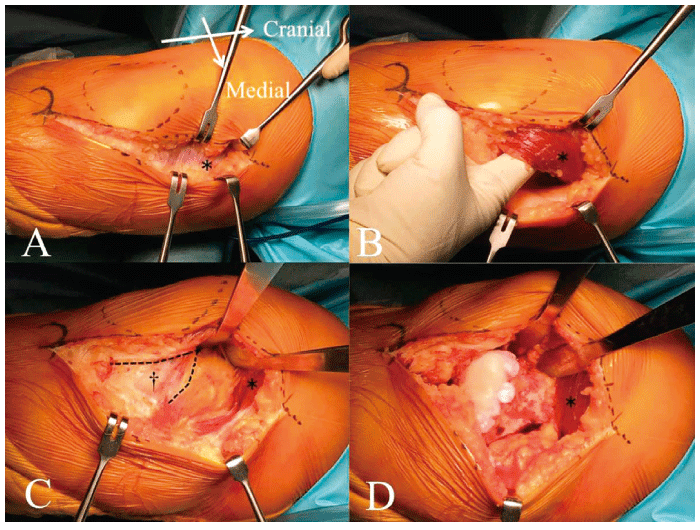
Fig. 1 Surgical exposure using the direct medial approach to the right knee
(A)A medial oblique incision of 14 cm is made from the medial aspect of the tibial tuberosity along the midpoint of the muscle belly of the vastus medialis obliquus muscle(*).(B)The muscle fibers are intentionally liftedup and lateralized within the sheath by the surgeon’s index finger.(C)The synovial layer of the suprapatellar pouch is visualized in a V-shaped flap(†).(D)The patella is lateralized with two narrow Homann retractors.
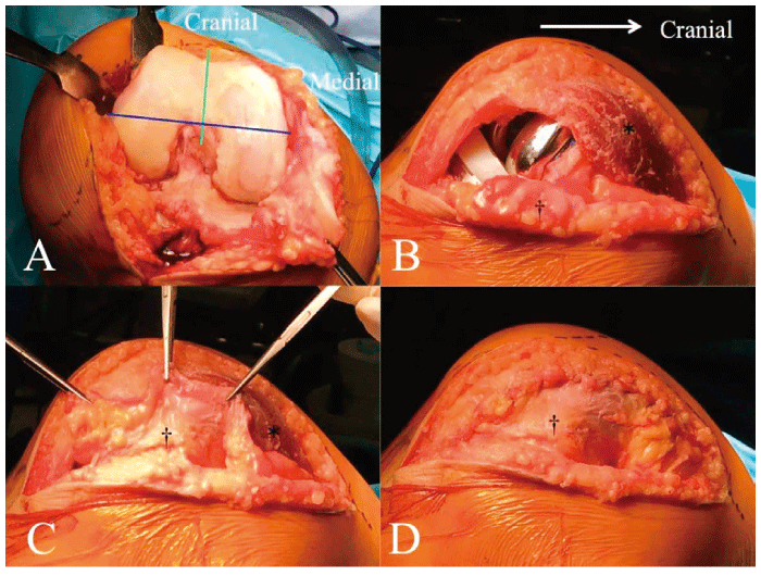
Fig. 2 Surgical exposure using the direct medial approach to the right knee(continued)
(A)The surgical epicondylar axis(blue line)and the Whiteside line(green line)in knee flexion.(B)The vastus medialis obliquus muscle(*)and the synovial flap(†)before closure.(C)The synovial flap is pulled up.(D)Reconstruction of the medial capsule with the synovial flap. Observe the complete covering of the vastus medialis muscle sheath.
The primary outcome was the surgical complication rate at three months. All adverse events(AEs), serious AEs(such as surgical site infection, cardiovascular events, and perioperative fracture), and AEs of special interest(revision surgery, iatrogenic severe sequelae, or death)were recorded. The secondary outcomes were surgical time, estimated blood loss by Gross’s formula [5], implant alignment, Knee Society score[6], and range of motion(ROM). Estimated total blood loss= Estimated blood volume×(preoperative hemoglobinpost operative-day one hemoglobin)/ preoperative hemoglobin+autologous blood transfusion volume+ allogeneic blood transfusion volume[5]. Implant alignment with the femoro-tibial angle(FTA), α angle, β angle, γ angle, and δ angle[7]were measured with Image J 1.45s(National Institute of Health, USA).
Baseline patient characteristics such as gender, age, diagnosis(osteoarthritis, rheumatoid arthritis, osteonecrosis), height, weight, body mass index, laterality, and physical status of the American Society of Anesthesiologists classification(ASA Physical Status Classification System, https://www.asahq.org/resources/ clinical-information/asa-physical-status-classificationsystem, accessed 25 Aug 2018)were recorded.
A consultant group(the first author was the primary surgeon)and a residents group(residents were the primary surgeons supervised by the first author)were compared with baseline patient characteristics and outcome, using Fisher’s exact probability test, Mann- Whitney U test, and Pearson χ2 test(JMP Pro 12.0, SAS, North Carolina). Predictive factors of outcome were determined using a step-wise multiple regression analysis.
A p-value<0.05 was considered significant.
The first author was the primary surgeon for 42 patients(consultant group), and 7 residents were the primary surgeons supervised by the first author for 58 patients(residents group). Baseline patient characteristics were not significantly different between the two groups(Table 1)
Forty-two percent of patients presented with AEs, and 10% with serious AEs, but no revision surgeries were necessary, and no iatrogenic severe sequelae or deaths occurred. Wound complications occurred in 20 patients, hypoxia in 9, disturbances of central nervous system in 5, fracture of any site in 3, urinary tract infection in 2, and tachycardia, urticaria, or whitlow in one each. The rate of all AEs was 36%(15 of 42 patients)for the consultant group and 47%(27 of 58 patients)for the residents group(p=0.310). Serious AEs included superficial surgical site infections in 6 patients(drug-resistant bacteria in five), intraoperative fracture in 2(crack of the tibial tuberosity and the femoral condyle), pulmonary embolism in one, and brain infarction in one. The rate of severe AEs was 12%(5 of 42 patients)for the consultant group and 9%(5 of 58 patients)for the residents group(p=0.738). All complications were completely resolved by conservative treatment.
Surgical time was significantly shorter for the consultant group than for the residents group(92±13 [mean±standard deviation] minutes versus 104±15 minutes, p=0.001). Estimated blood loss was similar for both groups(590±291 ml versus 485±240 ml, p=0.072). X-ray measurements were similar for both groups for postoperative FTA(176±3 degrees versus 176±3 degrees, p=0.620), α angle(97±2 degrees versus 97±2 degrees, p=0.267), β angle(89±2 degrees versus 89±2 degrees, p=0.969), and γ angle(8±3 degrees versus 6±3 degrees, p=0.080). The δ angle was one degree larger for the consultant group than for the residents group(89±2 degrees versus 88± 2 degrees, p=0.011). The postoperative knee score was similar for both groups(94±7 points versus 92± 7 points, p=0.051). The postoperative functional score was also similar for both groups(67±21 points versus 67±14 points, p=0.745). Postoperative total ROM was significantly larger for the consultant group than for the residents group(124±13 degrees versus 120±11 degrees, p=0.026). Postoperative maximum flexion also was significantly larger for the consultant group than for the residents group(125±13 degrees versus 121±11 degrees, p=0.030)
Multiple regression analysis revealed a predictive formula for postoperative total ROM;(postoperative total ROM[ degrees])=49.5+0.6×(postoperative pain score, none [50 points], mild [40 points], moderate[ 30 points])+0.4×(preoperative total ROM [degrees])+4.8×(surgeon, consultant [1 point], resident[ 0 point]), R=0.560, p=0.001. A scatter plot shows the correlation of preoperative and postoperative total ROM, and distribution of the consultant and residents groups(Fig. 3). A box-and-whisker plot shows better postoperative total ROM in patients without pain(Fig. 4).
Table. 1
Baseline patient characteristics in Consultant and Resident groups
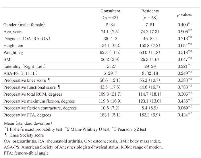
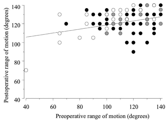
Fig. 3 Relationship of preoperative and postoperative ROM between consultant and residents
(Postoperative ROM)=97+0.2×(preoperative ROM), R=0.361, p=0.001, simple regression analysis. White dots: consultant group, black dots: residents group, and gray dots: overlapping of consultant and residents groups.
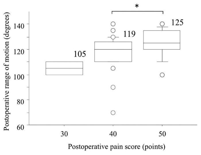
Fig. 4 Comparison of postoperative pain score and ROM
Box-and-whisker plot shows the mean ROM and standard deviation. Mann-Whitney U test followed by Bonferroni correction. *p=0.013
The direct medial approach(DMA)is a novel surgical technique for primary TKA, characterized by a medial oblique skin incision and preservation of a V-shaped synovial flap at the medial deep layer of the joint capsule. We modified the skin incision from the original subvastus approach[1]that used an anterior straight line, to a medial oblique line. The new incision has several advantages. First, it can directly expose the vastus medialis muscle and the medial aspect of the capsule. There is little postoperative swelling or tenderness around the anterior aspect of the patella. Second, skin tension with the knee flexed is less on the medial side using this approach than it is with an anterior straight line. The oblique incision works in favor of wound healing and deep flexion(Fig. 5). Serious AEs occurred in 10% of the patients, and the values of residents were similar to those of the consultant group. Yohe et al. [8]reported 19.3% of minor complications and 3.5% of major complications after TKA in octogenarians and nonagenarians. Kim et al.[9]reported 13% of complications in minimally invasive technique and 6% of complications in standard technique. Therefore, complication rate would be influenced by the definition of complications, patient’s characteristics, surgical approach, surgeon’s learning curve and so on. In this study, all the complications completely resolved without revision surgery or any sequelae by conservative treatment. We suggest that DMA is a safe technique and provides a sufficient exposure for TKA.
The medial para-patellar approach is a gold standard technique for varus-type knee OA for both primary and revision TKA. It provides excellent exposure and has a very low incidence of complications. However, this approach results in the loss of vascular supply to the patella, and denervation and muscle weakness of the quadriceps [10]by separating the VMO from the patella and the rectus femoris muscles intramuscularly. The mid-vastus approach also causes deterioration of the vastus medialis muscle. An historical cohort of patients subjected to the mid-vastus approach with the same implant and same surgeon[4]averaged 119.3 degrees of ROM postoperatively, 4.7 degrees less than the consultant group in this study. The lateral para-patellar approach is more appropriate in valgus-type knee OA for TKA. The subvastus approach is the most anatomical procedure to expose the knee joint when compared to other conventional approaches. The subvastus approach provides better pain relief with lower opioid use and quick recovery with earlier straight leg raising and greater knee flexion[11,12]. Compared with the medial para-patellar approach, patellar tracking is more natural with the subvastus approach due to preservation of the extensor mechanism[12,13]. This study supports these previous opinions by showing better postoperative total ROM in patients without pain(Fig. 4)
A proximal landmark of the mid-point of the muscle belly of the vastus medialis was applied in DMA, because the muscular branch of the descending genicular artery(DGA)runs along the medial edge of the muscle [3]. It is true that it is technically demanding using the subvastus approach to obtain sufficient exposure of the surgical field, especially to evert the patella[11,14]. To improve the exposure, the skin incision must be extended proximally because the distal end is limited by the tibial tuberosity. For proximal extension, attention must be paid to the vascular anatomy. The DGA, the final branch of the femoral artery, is at risk of damage, even though it is protected by the intermuscular fascia and the adductor hiatus. The surgeon must be aware of the anatomical features of the DGA, which has anatomical variations in branching patterns[15]. Fauré et al stated there were two patients with symptomatic medial-thigh hematomas caused by damage to the DGA [10]. Some cadaveric studies suggest that the safe zone between the DGA and the medial joint line is 12.8- 14.5cm[16,17]. Another cadaveric study reported that the mean distance from the tibial tuberosity to the DGA was 15.5 cm[3]. In fact, no vascular injuries were observed in the current study. We suggest that an oblique medial skin incision within 14 cm of the tibial tuberosity along the mid-point of the muscle belly of the VMO is a safe zone for DMA to avoid DGA damage.
Intraoperative patellar eversion was a reliable indicator of sufficient exposure with or without patellar replacement. It is true that lateral exposure is technically demanding in DMA. Exposure can be facilitated by resection of the dense connective tissue between the suprapatellar pouch and the vastus intermedius muscle. In knee hyper-extension, the patella is pulled medially and gradually everted. The lateral capsule is minimally released at the lateral aspect of the patella from inside the joint with electrocautery. Excessive lateral release increases the incidence of wound complications [18]. In a severely stiff knee, eversion of the patella becomes more difficult and requires release of the lateral patellofemoral ligament and removal of osteophytes from the lateral femoral condyle and patellar edge.
There are several limitations to this preliminary report. First, this was a technical note of a novel surgical procedure and perioperative safety. A follow-up period of three months is short, but in general the advantage of minimally invasive surgery is a quick recovery within the first three months postoperatively. Longer follow-up is needed to provide more confidence in the long-term positive outcomes when using this technique. Second, this study only showed a prospective cohort of primary TKA in“ standard” patients. Relative contraindications of DMA may be valgus deformity, severe contracture, obesity, previous major knee arthrotomy or revision TKA. In these conditions, eversion of the patella may be much more difficult with poor visualization.
In conclusion, the direct medial approach is a novel and safe technique for primary TKA over the first three months postoperatively. It uses a medial oblique skin incision and preserves a synovial flap at the medial deep layer of the joint capsule.

Fig. 5 Comparison of the operative scar after two different incisions
An 80-year-old woman underwent arthroplasty of her right knee using a mid-vastus approach via a midline incision, followed by the direct medial approach with a medial oblique incision on her left knee. Anteroposterior view in knee extension(A). Distal view of the right(B)and left knee(C)in flexion. Maximum flexion is 115 degrees in the right(D)and 135 degrees in the left knee (E). Proximal view of the right(F)and left knees(G)in flexion.
JN, KS, TS, SH, and SO1 analyzed and interpreted the patient data regarding the clinical outcome. JN, TN, YE, and KI measured the X-rays. JN, TA, YS and SO2 planed the study design. KS and TS made substantial contributions to conception. JN wrote the manuscript. SH, TA, TN, and YE revised it critically for important intellectual content. JN and KS equally contributed the article as the first author. All authors read and approved the final manuscript.
Source of funding
The first author(JN)was supported by JSPS KAKENHI grant number 17K10954 and Takeda Science Foundation.
None.
Address correspondence to Dr. Junichi Nakamura.
Department of Orthopaedic Surgery, Graduate School of Medicine, Chiba University, 1-8-1, Inohana, Chuou-ku, Chiba 260-8670, Japan.
Phone: +81-43-226-2117. Fax: +81-43-226-2116.
E-mail:njonedr@chiba-u.jp
