


Chiba Medical J. 96E:27-31, 2020
doi:10.20776/S03035476-96E-1-P27
[ Case Report ]
Nao Baba1) , Hiroshi Ishikawa1) , Tatsuya Kobayashi1),
Ayumu Matsuoka1) , Michiyo Kambe2) , and Makio Shozu1)
1) Department of Reproductive Medicine, Graduate School of Medicine, Chiba University, Chiba 260-8670 .
2) Department of Diagnostic Pathology, Chiba University Hospital, Chiba 260-8677 .
(Received September 1, 2019, Accepted October 23, 2019, Published February 10, 2020.)
The signs of granulosa cell tumors (GCTs) arising from the ovary depend on the types of hormones produced by the tumor. Estrogens cause continuous uterine bleeding, whereas inhibins are responsible for amenorrhea. We describe an unusual case of GCT in which the tumor-related manifestations changed over 9 years, from initial secondary amenorrhea to continuous uterine bleeding. Magnetic resonance imaging revealed a suspected ovarian tumor, and bilateral adnexectomy was performed. The preoperative serum hormone levels (luteinizing hormone, 0.5 mIU/mL; follicle-stimulating hormone, <0.05 mIU/mL; estradiol, 279 pg/mL) returned to the normal menopausal range after tumor resection (11.43 mIU/ mL, 23.80 mIU/mL, and <10 pg/mL, respectively) . The preoperative serum inhibin B levels were high, and the tumor tissue was diffusely immunostained with inhibin α. The woman had never experienced estrogen-deficiency symptoms. Therefore, we believe that the tumor produced inhibins initially, which led to hypogonadotropic amenorrhea that manifested as apparent early menopause, and estrogens subsequently, which caused continuous uterine bleeding over the years.
Granulosa cell tumor, menopause, amenorrhea, inhibins, ovary, estrogen
Granulosa cell tumors (GCTs) are sex cordstromal cell tumors that arise from the ovaries and produce different types of hormones, including estrogens, androgens, and inhibins that cause specific and pathognomonic symptoms. In menopausal women recurrent abnormal uterine bleeding is the most common sign of estrogen-producing GCTs[1]. Persistent amenorrhea, although rare, is another sign of GCTs and is caused by inhibins[2]. Virilization (hirsutism and acne) can be caused by androgens released from GCTs[3]
Women with GCTs exhibit any of these manifestations of tumor-producing hormones[4]. However, it is rare for GCTs to produce metachronous multiple manifestations. We report the case of a 50-year-old woman with GCT whose manifestations changed over a period of 9 years: persistent amenorrhea for 9 years, which was thought to be early menopause, followed by continuous uterine bleeding for 1 year.
A 50-year-old nulliparous woman who was obese (height, 154 cm; weight, 81 kg; body mass index, 34) presented with lower abdominal pain and a 1-year history of continuous uterine bleeding. Menarche had occurred at 13 years of age, and her menstrual cycles were regular until the age of 40, when amenorrhea developed. She reported never having experienced menopause-related symptoms, such as hot flashes, moodiness, or episodic sweating. A tender lower abdominal tumor (22 × 18 × 8 cm) was found on magnetic resonance imaging (MRI) . The height of uterine fundus was 10.8 cm at the long axis, and the endometrium was 8 mm thick (normal for the proliferative phases) . Trace amounts of dark red blood were present in her vagina; however, characteristic postmenopausal atrophic changes were not apparent in her vulva or vagina. Results of cervical and endometrial cytology tests were negative for malignancy.
An endometrial biopsy revealed endometrial tissue in mixed proliferative and secretory phases, which indicated a continuous estrogenic effect on the endometrium. MRI revealed a right ovarian multilocular, cystic tumor comprising a solid part with low- and highintensity areas on T2-weighted images (Fig. 1) . A few small cysts were also noted in the left ovary. The MRI findings were consistent with the diagnosis of GCT. The left ovary was 2.5 × 2.0 × 1.8 cm in size. Blood examination revealed normocytic anemia (red blood cell count, 25.3 × 10⁶/mL; hemoglobin, 6.7 g/dL; hematocrit, 21.3%; mean cell volume of red blood cells, 84.9 fL) . Serum hormone levels-low gonadotropins and high estradiol-were not characteristic of menopause (Table 1) . Inhibins are heterodimers consisting of α, βA, and βB subunits with a disulfide bond. We identified elevated levels of serum inhibin B (754.1 pg/mL; normal range, 10-200 pg/mL) by using the RayBio® Human/Mouse/Rat Inhibin B (Beta B subunit) Enzyme Immunoassay Kit (RayBiotech, Norcross, GA, USA)[5].
We performed total hysterectomy, bilateral adnexectomy, and partial omentectomy. The tumor capsule was found to be partly ruptured. Results of peritoneal cytologic tests were negative for malignancy. Pathological study of the resected tumor demonstrated the presence of an adult-type GCT (stage pT1c2 (IC2) , according to the International Federation of Gynecology and Obstetrics[FIGO]classification of ovarian cancer[6]) with Call-Exner bodies and coffee bean-shaped nuclei. Immunostained tumor cells showed diffuse expression of inhibin α (Fig.2) . With genomic DNA extracted from frozen tumor tissue, we performed direct sequencing of the FOXL2 gene. The tumor tissues were heterozygous for a missense mutation at c.402C>G (p.C134W) , a frequent observation in adult-type GCT[7].
After the surgery, serum gonadotropin levels increased to the postmenopausal range, and the serum estrogen levels became undetectable (Table 1) . The serum inhibin B levels were increased slightly on day 1 after surgery (903.5 pg/mL) , and were decreased on day 7 (647.4 pg/mL) . We administered adjuvant chemotherapy in six cycles of paclitaxel (175 mg/m2) and carboplatin (area under the curve, 6) every 3 week, in accordance with the standard regimen for women with ovarian cancer. No recurrence of the tumor has been observed for five years after the surgery.
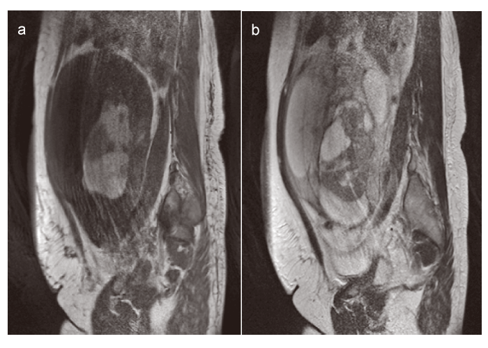
Fig.1 Magnetic resonance imaging findings of the tumor
(a) Sagittal T1-weighted image. Maximum tumor diameter was 22 cm. Bleeding area showed high intensity.
(b) Sagittal T2-weighted image. The multilocular cystic mass included low- and high-intensity areas. A solid part was recognized in the cystic tumor.
Table.1
Serum gonadotropin and sex steroid levels before and after surgery.
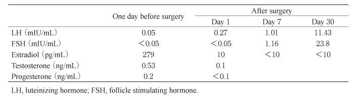
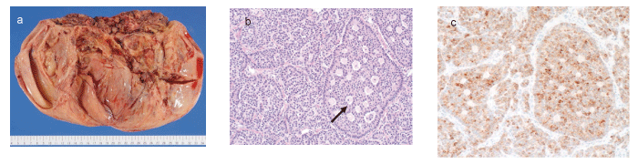
Fig.2 Pathological findings of the ovarian tumor
(a) Macroscopic findings of the tumor. The surface and lumen of the cyst were smooth. A necrotic lesion with massive bleeding was observed in the solid portion. (b) Microscopic findings of the tumor at low magnification (×40) . Call-Exner bodies (arrows) was observed in the lumen of the cyst. (c) Immunohistochemistry of the tumor for inhibin α. We observed diffuse inhibin α expression around the lumen of the gland portion.
To our knowledge, no case of GCT like oursmanifesting with amenorrhea for 9 years, followed by continuous abnormal uterine bleeding-has been reported. We believe that both the persistent amenorrhea, which was assumed to be early menopause, and continuous uterine bleeding were associated with a transition in the expression of the major tumor-induced hormones, from inhibin to estrogen. The prolonged amenorrhea may have resulted from hypogonadotropic anovulation, and the continuous uterine bleeding may have represented endometrial breakthrough caused by hyperestrogenemia.
Secondary amenorrhea is one of the signs of an inhibin-producing GCT. Excess inhibins secreted from the GCT may suppress pituitary secretion of folliclestimulating hormone (FSH) , which in turn suppresses ovulation and results in secondary amenorrhea. Such hypogonadotropic amenorrhea in a woman 40 years of older is assumed to represent menopause. Our patient exhibited extremely low levels of serum FSH and high levels of serum inhibin B while uterine bleeding continued; after resection of GCT, the FSH levels returned to menopausal levels. Therefore, we concluded that her secondary amenorrhea, which is an apparent early menopause, was caused by hypogonadotropic hypogonadism.
In a previous report of GCT, a woman presented with menopause at 44 years of age[8]; at the age of 48, this patient complained of atypical vaginal bleeding, and laboratory testing revealed undetectable FSH levels in association with a high estradiol level, as in our patient. In another reported case of GCT, a woman presented with menopause at the age of 43, followed by postmenopausal mild hirsutism and hyperandrogenemia[9]. Resumption of menstruation in women with GCTs who presented with secondary amenorrhea has also been reported.
Although the mechanisms of menstrual cessation in women with hypogonadotropic hypogonadism differ from those in menopausal women, both conditions lead to hypoestrogenemia, which causes estrogendeficiency symptoms (including neurovegetative and psychoemotional disorders, urogenital abnormalities, and vasomotor symptoms) . However, our patient was not aware of any estrogen-deficiency symptoms. The absence of estrogen-deficiency symptoms may indicate that her serum estradiol levels were maintained by the GCT-producing estrogen. The levels of serum inhibins and estradiol are not correlated in women with GCTs that produce inhibins[2,10]. Serum estradiol levels are affected by both the pituitary-gonadal axis and tumorproducing estrogen in women with GCTs. Although tumor-producing inhibins may inhibit pituitary FSH secretion, the serum levels of estradiol do not decrease to the menopausal range. In contrast, women with ovarian tumors that produce only inhibin B present with hypogonadotropic amenorrhea and estrogen-deficiency symptoms. A woman with such an ovarian tumor who presented with secondary amenorrhea and hot flushes has been reported[5].
Heterozygosity for a missense mutation of the FOXL2 gene at c.402C>G (p.C134W) was identified in our patient. Forkhead box L2 protein is a key transcription factor for the proliferation and the differentiation of granulosa cells in the ovary, and the somatic missense point mutation in the FOXL2 gene is characteristic in adult-type GCT. Although the role of this mutation in the pathogenesis of adult-type GCT remain uncertain, its presence leads to increased proliferation and longer survival of granulosa cells and promotes hormonal changes[11]. In addition, this mutation is preserved in recurrent GCTs, which suggests that it is oncogenic and critical for the tumorigenesis of adult-type GCT[12].
In our patient, the serum inhibin B levels, which were high before surgery, increased slightly on day 1 after surgery and were decreased on day 7 after the surgery; however, all of levels were abnormally high. Although the precise half-life of inhibin B in human remains unknown, the serum half-lives of the transforming growth factor β family, to which inhibins belong, are short[13]; therefore, metabolic clearance of inhibin B might have been impaired in our patient. In addition, serum levels of inhibins remained high during the continuous uterine bleeding, and this may have caused the secondary amenorrhea to last for 9 years.
In conclusion, women with GCTs present with specific signs associated with the hormones produced by the tumors. Changes in the main sign may result from a change in the dominant hormone produced by a GCT. Both serum hormone levels and the associated symptoms are helpful for the identification of GCTs.
N. B. corrected patient’s data, made figures and a table, and prepared the manuscript. H.I. summarized the data, revised the figures and a table, and prepared the manuscript. T.K. measured serum inhibin levels. A.M. got written consent from the patient and revised the manuscript. M.K. prepared pathological findings. M.S. revised the manuscript.
This work was supported by the JMWH Bayer grant (2018) . The authors would like to thank Enago (www. Enago.jp) for the English language review.
The authors declare that there is no conflict of interest regarding the publication of this manuscript.
Address correspondence to Dr. Hiroshi Ishikawa.
Department of Reproductive Medicine, Graduate School of
Medicine, Chiba University, 1-8-1, Inohana, Chuou-ku, Chiba
260-8670, Japan.
Phone: +81-43-226-2121. Fax: +81-43-226-2122.
E-mail:ishikawa@chiba-u.jp
