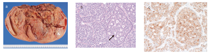
Fig.2 Pathological findings of the ovarian tumor
(a) Macroscopic findings of the tumor. The surface and lumen of the cyst were smooth. A necrotic lesion with massive bleeding was observed in the solid portion. (b) Microscopic findings of the tumor at low magnification (×40) . Call-Exner bodies (arrows) was observed in the lumen of the cyst. (c) Immunohistochemistry of the tumor for inhibin α. We observed diffuse inhibin α expression around the lumen of the gland portion.