


Chiba Medical J. 96E:47-54, 2020
doi:10.20776/S03035476-96E-2-P47
[ Original Article ]
Seiji Kimura1,2), Satoshi Yamaguchi2,3), Aya Sadamasu1,2),
Yuya Ogawa1,2), Yoshimasa Ono1,2), Shotaro Watanabe1,2),
Ryuichiro Akagi2), Seiji Ohtori2), and Takahisa Sasho1,2)
1) Musculoskeletal disease and pain, Preventive Medical Sciences, Chiba University, Chiba 260-8670.
2) Department of Orthopaedic Surgery, Graduate School of Medicine, Chiba University, Chiba 260-8670.
3) Collage of Liberal Arts and Sciences, Chiba University, Chiba 263-8522.
(Received November 9, 2019, Accepted November 27, 2019, Published April 10, 2020.)
The objective of this research is to mechanically and histologically evaluate the capacity of different PRP concentrations to promote tendon healing.
60 New Zealand white rabbits were used. In 20 rabbits (L-PRP group), 1.0 mL of lowconcentration PRP was applied to the Achilles tendon tear transection site on both hindlimbs of each animal. The other 20 rabbits (H-PRP group), 1.0 mL of high-concentration PRP and in the remaining 20 rabbits (S group), 1.0 mL of saline were applied. Ten rabbits from each group were slaughtered at 4 and 8 weeks following the transection surgery, with subsequent histological evaluation of the right legs, and mechanical evaluation of the left legs.
Platelet counts were 3.8 times higher for L-PRP and 12.8 times higher for H-PRP than for whole blood. The semi-quantitative Bonar scale (0 is normal) decreased significantly from the S to L-PRP to H-PRP groups (P=0.03). The values for ultimate load, ultimate stress, elastic modulus, and stiffness increased from the S to L-PRP to H-PRP groups. A significant difference was observed between the groups for stiffness (P=0.03)
Our results showed that PRP preparations with a high concentration of platelets were superior in the promotion of histological and mechanical healing of the Achilles tendon.
platelet-rich plasma (PRP), Achilles tendon, rabbit, tendon healing, tendon rupture
Achilles tendon rupture is a common sports injury [1]. It takes time for the tendon to heal after such an injury, and often 6-9 months are needed before the athlete can resume sports activity[2,3]. Several methods to accelerate tendon healing are currently being tested, including treatment with atelocollagen and boric acid[4,5]. However, no definitive method exists, save early exercise therapy[6]. Platelet rich plasma (PRP) is blood plasma enriched in platelets obtained by centrifugally separating blood and contains a high concentration of growth factors. Platelets contain over 300 physiologically active proteins, including vascular endothelial growth factor (VEGF), fibroblast growth factor (FGF), platelet-derived growth factor (PDGF), and transforming growth factor beta (TGFb), and these factors can potentially encourage tendon healing [7-10]. However, the research results in animals and humans regarding the effects of PRP on Achilles tendon healing are inconsistent [8-11]. It is speculated that this may be due to a lack of standardized quality for PRP preparations, including the presence of white blood cells in PRP, the number of centrifugal separations, and varying enrichment factors. DeLong advocated PAW classification[12], which is a classification based on PRP components and activation methods, but very few studies have investigated the effects of various concentrations of PRP on tendon healing. There is no consensus on the most appropriate platelet concentration in PRP.
The objective of this research is to mechanically and histologically evaluate the capacity of different PRP concentrations to promote tendon healing.
Animals
All animal experimentation protocols were approved by the Ethics Committee of this institution and are in accordance with the Animal Experimentation Management and Use Guidelines of the National Institutes of Health.
Mature New Zealand white rabbits (age, 24-26 weeks; weight, 2.8-3.6 kg) were used in this study. All rabbits were randomly grouped into three groups. In 20 rabbits (L-PRP group, 40 hindlimbs), 1.0 mL of lowconcentration PRP was applied to the Achilles tendon tear transection site as described in the next section. The other 20 rabbits (H-PRP group, 40 hindlimbs), 1.0 mL of high-concentration PRP was applied to the tear transection site. In the remaining 20 rabbits (S group, 40 hindlimbs), 1.0 mL of physiological saline. Finally, 6 rabbits (C group, 12 hindlimbs) served as controls, and these animals did not have transected Achilles tendons. The number of animal subjects and amounts of administered PRP were based on previous reports[13-16]. Ten rabbits from each group were slaughtered at 4 and 8 weeks following the transection surgery, with subsequent histological evaluation of the right legs, and mechanical evaluation of the left legs.
PRP Preparation
PRP is defined as the plasma layer fraction rich in platelets formed by centrifuging blood[17]. Rabbits were intramuscularly injected with a mixture of medetomidine hydrochloride (0.5 mg/kg, Domitor, Nippon Zenyaku Kogyo, Japan), midazolam (2.0 mg/kg, Dormicum, Astellas Pharma, Japan), and butorphanol tartrate (0.5 mg/kg, Betorfal, Meiji Seika Pharma, Japan) to induce anesthesia. Blood (highest possible volume) was drawn from the pericardium with an 18-gauge needle. Immediately after collection, the blood was transferred into tubes containing anticoagulants. The collected blood was utilized to prepare low-concentration (10 mL) PRP (L-PRP), high-concentration (40 mL) PRP (H-PRP), and 2 mL was reserved for blood counts. The PRP was prepared by centrifugation (once) using a commercial platelet separation kit (MyCells Autologous Platelet Preparation System, Kaylight, Ramat-Hasharon, Israel). In accordance with previous reports, the centrifugation time was 10 minutes at 1500 G. L-PRP (2.5 mL) was prepared from 10 mL of whole blood, and H-PRP (2.5 mL) was prepared from 40 mL of whole blood[18]. Platelet activation, was induced in PRP preparations by treatment with 0.2% CaCl2 to cause gelation[19]. We administered 1.0 mL of PRP to each hindlimb in the PRP group. Whole blood and any remaining PRP were used to calculate platelet, white blood cell, and red blood cell counts using a hemocytometer (MEK-6358 Celltac α, Nihon Kohden, Tokyo, Japan).
Achilles Tendon Transection Model
Rabbits were anesthetized as described above. After creating a vertical incision in the skin, the paratenon was split vertically, exposing the Achilles tendon. The Achilles tendon was horizontally transected approximately 2 cm from its calcaneal attachment point. The plantar fascia was untouched and allowed to serve as an internal splint [13]. Subsequently, the L-PRP and H-PRP groups were treated with 1.0 mL of PRP (of the appropriate concentration) at the tendon rupture site. The S group, was treated with the same volume of physiological saline instead of PRP. After administration of either PRP or saline, the paratenon and the animal’s skin were sutured closed with 4-0 nylon, and this procedure was carried out on both hindlimbs of each animal. After surgery, animals were returned to their cages and allowed to move about freely.
Histological Evaluation
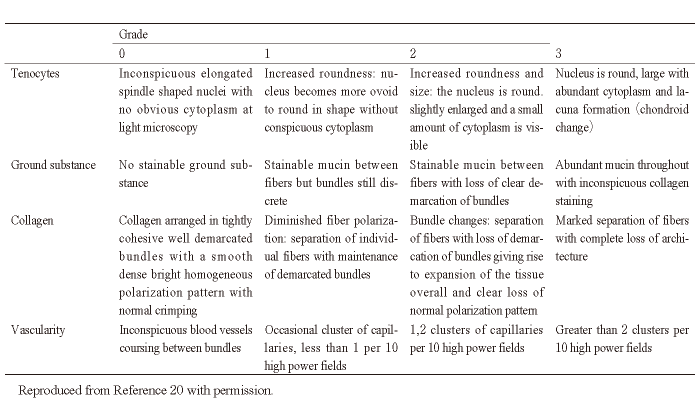
Histological evaluations were conducted at 4 and 8 weeks after treatment with PRP or physiological saline. The right Achilles tendon was removed and separated from the surrounding tissue. Samples were fixed in a buffered 10% formalin solution and embedded in paraffin blocks. Sections were cut at 4 mm intervals along the sagittal plane of the Achilles tendon. Two paraffin sections were created at the center of the healing area for each tendon and one was stained with hematoxylin eosin, while the other was stained with Masson Trichrome. We performed histopathological analysis of the Achilles tendon by utilizing the semiquantitative Bonar scale[20]. This scale consists of four subscales, each scored 0, 1, 2, or 3, where 0 is normal and 3 is maximally abnormal. Scale one evaluates the presence of tendon cell morphology and proliferation, two evaluates the presence or absence of the intercellular matrix, three the characteristics of the collagen bundle, and four, angiogenesis (Table 1). The overall Bonar scales were calculated as the sum of each subscale score. The area with the most marked pathological change, at the center of the tendon healing area on each slide, was selected for evaluation. Each histological analysis was performed separately by 2 different orthopedic surgeons who were blinded to the group allocations with 5 years of experience each, and the mean Bonar scale score was designated as the final value. The interclass correlation coefficient between observer measurements was 0.85.
Mechanical Evaluation
Mechanical evaluations were conducted at 4 and 8 weeks following treatment with PRP or physiological saline. The left Achilles tendon of each animal was extracted alongside the calcaneus and gastrocnemius soleus. Using specially designed equipment, clamps were attached to the calcaneus and the gastrocnemius soleus (40 mm away from the attachment of the Achilles tendon to the calcaneus), and placed 40 mm apart from each other (Autograph AG-X mechanical testing apparatus, Shimadzu Corp., Kyoto, Japan). Prior to testing, samples were preloaded with 5 N of force at 24±1℃ for 5 minutes to align the collagen fibers[21,22]. Following the preloading, samples were pulled at a rate of 10 mm/min, and the ultimate load (N), ultimate stress (MPa), elastic modulus (MPa), and stiffness (N/mm) were determined [23,24]. Elastic modulus was calculated to correspond to 20% to 80% of ultimate stress [25]. Stiffness was calculated as the slope of the load-displacement curve corresponding to 20% to 80% of the ultimate load[25]
Statistical Analysis
The white blood cell and platelet counts of PRP preparations were expressed as the median (25th-75th percentile), histological and mechanical assessments were expressed as mean ± standard deviation. The white blood cell and platelet counts of PRP preparations were compared using the Kruskal-Wallis test and the Tukey-Kramer test was used for post-hoc analysis.
Changes over time, and the differences in treatment effects for each evaluation metric were calculated using a linear mixed-effects model. The number of weeks after the surgery and evaluation metrics were set as dependent variables, the random effect was set as each animal, and the correlation between variables was investigated. A linear mixed-effects model was used to calculate statistics with the commercial SAS software package (SAS Institute, Sally, NC, USA), and BellCurve for Excel (Social Survey Research Information Co., Ltd, Japan) was utilized for other tests. The P-value threshold for statistical significance was set to 0.05.
Bioethical approval
All experimental procedures in this study were approved by the Animal Experiment Committee of Chiba University (Approval No. 30-330), and were conducted in accordance with the Guidelines for Proper Conduct of Animal Experiments of the Science Council of Japan.
Table. 1 Bonar Scale
Bioethical approval
All experimental procedures in this study were approved by the Animal Experiment Committee of Chiba University (Approval No. 30-330), and were conducted in accordance with the Guidelines for Proper Conduct of Animal Experiments of the Science Council of Japan.
Platelet counts were 84.2 (56.4-99.9) (x 104/ μl) for L-PRP, and 284.2 (192.1-328.1) (x 104/μl) for H-PRP, and both were significantly higher than the platelet counts for whole blood[ 22.1 (17.5-25.0) (x 104/μl)] (P<0.001); specifically, 3.8 and 12.8 times higher, respectively. White blood cell counts were 2010 (1540-3200) (/μl) for L-PRP, and 2290 (1530-3380) (/μl) for H-PRP; both figures were lower than the count for whole blood[ 3085 (1510-4510) (/μl)], but no significant difference was observed between the three groups (P=0.51).
Histological Evaluation
Tendon cell morphology, intracellular matrix volume, collagen arrangement, and blood vessel numbers were evaluated. As tendon healing progressed, the number and size of tendon cells decreased, as did the size of the intracellular matrix. Collagen fibers began to be arranged in parallel along their long axes. The number of blood vessels decreased (Fig. 1). Bonar scale values decreased significantly from the 4th to the 8th week (P=0.01). Bonar scales decreased from the S to L-PRP to H-PRP groups (Table 2), and a significant difference in treatment effect was observed between the groups (P=0.03).
Mechanical Evaluation
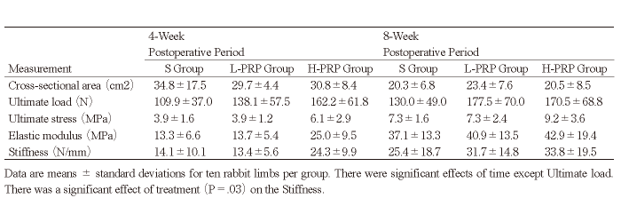
The transected cross-sectional area, ultimate load (N), ultimate stress (MPa), elastic modulus (MPa), and stiffness (N/mm) were evaluated. The transected cross-sectional area, ultimate load (N), ultimate stress (MPa), elastic modulus (MPa), and stiffness (N/mm) for the control group were 20.4±4.2 (cm2), 215.4± 71.1 (N), 10.4±3.4 (MPa), 106.9±29.3 (MPa), and 71.8±17.5 (N/mm), respectively. The transected crosssectional area decreased significantly from the 4th to the 8th week in each group (P<0.001). The ultimate stress, elastic modulus, and stiffness increased significantly from the 4th to the 8th week. The values for ultimate load, ultimate stress, elastic modulus, and stiffness increased from the S to L-PRP to H-PRP groups (Table 3). A significant difference in treatment effect was only observed between the groups for stiffness (P=0.03).
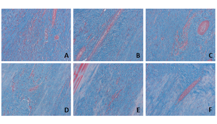
Fig. 1 Photomicrographs of the Achilles tendon. A-F, Photomicrographs of the Achilles tendon (Masson trichrome stain; original magnification, ×100).
Representative images from, A-C, 4-week postoperative period (A: C group, B: L-PRP group, C: H-PRP group) and, D-F, 8-week postoperative period (D: C group, E: L-PRP group, F: H-PRP group) are shown.
Table 2 Bonar Scales for the Achilles Tendon
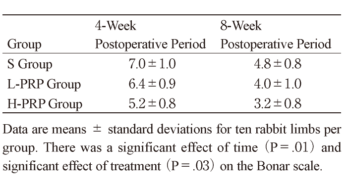
Table 3 Mechanical Assessment for the Achilles Tendon
In this study, Bonar scale values were highest for the S group, followed by the L-PRP group, and were lowest in the H-PRP group, indicating that histological evidence for promotion of tendon healing was most pronounced for high-concentration PRP preparations. The mechanical analyses also revealed that promotion of tendon healing evidence was highest in the H-PRP group, followed by the L-PRP group, and was lowest for the S group. A significant difference was observed for stiffness.
PRP is plasma enriched in platelets, and contains a high concentration of growth factors, which can potentially promote tendon healing. However, a consensus on the most appropriate concentration of platelets has not been reached. The PAW classification system of PRP is based on 3 components: (1) the absolute number of Platelets, (2) the manner in which platelet Activation occurs, and (3) the presence or absence of White cells[12]. Platelet concentration categorized as follows: P1, less than or equal to baseline levels; P2, greater than baseline levels to 750,000 platelets/μL; P3, greater than 750,000 to 1,250,000 platelets/μL; and P4, greater than 1,250,000 platelets/μL. In this classification, L-PRP in this study is classified as P3, and H-PRP is classified as P4. Ours is the first report on the tendon-healing-promotion effects of different concentrations of PRP, in a rabbit model of Achilles tendon transection. Mastrangelo et al. reported no significant difference between platelet concentrations of 3x and 5x in the mechanical healing of a sutured ACL in animal models[26]. However, the difference in concentration between the two conditions was rather small. We used concentrations of 3.8x and 12.8x, and found that stiffness was significantly higher for the high-concentration group, likely because our PRP concentrations differed by a wider margin. While a significant difference in treatment effect between the three groups was only observed for stiffness, stiffness has been reported as the most clinically important structural characteristic for tendons and ligaments[27-29], and we believe that PRP concentration had an effect on its capability to promote mechanical healing of the affected tendon.
In this study, high-concentration PRP preparations were better for the promotion of histological and mechanical healing of affected tendons. Platelet and growth factor concentrations are reportedly proportional to one another [30,31]. This suggests that the H-PRP preparations in our study contained a higher level of growth factors, possibly enhancing the promotion of tendon healing. Increasing the number of centrifugations, speed, and running time can increase the enrichment factor (concentration) of PRP preparations [12], but these more vigorous extraction procedures can damage platelets, and cause depletion of necessary growth factors for tissue healing. In this study, we minimized the effect of the extraction process on the integrity of platelets in the low- and high-concentration PRP preparations by not altering the centrifugation parameters, and instead using more blood for the H-PRP preparation to yield a higher number of platelets.
Our research has several limitations.
First, the number of tendons we examined was relatively small (10 per group, per timepoint). While the number of animals sacrificed for the purpose of this research should be kept to a minimum, further investigations that make use of more samples would strengthen our findings.
Second, we did not evaluate the growth factor concentrations in our PRP preparations. However, even though we did not directly measure the growth factor concentrations in the H-PRP and L-PRP preparations, previous reports showed that platelet and growth factor concentrations were proportional to each other, and the extraction processes used to prepare both types of PRP used in our study were the same as previously reported. Thus, we can expect that the growth factor concentration was higher in the H-PRP than in the L-PRP preparations.
Third, we administered physiological saline to the surgery site in our control group, instead of PRP. Lyras et al. [33]also injected their study control group with physiological saline; however, it is possible that saline has an effect on the tendon healing process. In addition, since we transected Achilles tendons and administered PRP and saline without taking time, local bleeding may affect the results.
Fourth, in our statistical analyses, we treated the two Achilles tendons from each rabbit as independent from one another. However, it is quite possible that the healing processes in two different tendons from a single rabbit are related. However, from an animal welfare standpoint, keeping the number of rabbits used in our study to a minimum required using the tendons on both legs.
Fifth, a rabbit model of Achilles tendon rupture will differ from a clinical model of Achilles tendon rupture in humans. Thus, it may well be the case that our findings with respect to PRP concentration do not directly translate to the clinical treatment for a ruptured Achilles tendon in human patients. However, the main objective of this research was to compare the effect of different concentrations of PRP preparations. Furthermore, we must keep in mind that the mechanical and histological analysis of Achilles tendon rupture in human subjects is ethically questionable and technically difficult.
In conclusion, We evaluated the potential of different PRP concentration preparations to promote tendon healing in a rabbit Achilles tendon transection model. Our results showed that PRP preparations with a high concentration of platelets were superior in the promotion of histological and mechanical healing of the Achilles tendon.
Guarantor of integrity of the entire study: SO, TS. Study concepts and design: All authors. Clinical studies: SK, YO. Manuscript preparations: SK. Manuscript editing: TS.
The authors declare that they have no conflicts of interest, either financial or non financial, with the contents of this article.
Address correspondence to Dr. Takahisa Sasho.
Musculoskeletal disease and pain, Preventive Medical Sciences,
Chiba University, 1-8-1, Inohana, Chuou-ku, Chiba 260-8670,
Japan.
Phone: +81-43-226-2961. Fax: +81-43-224-5124.
E-mail:sasho@faculty.chiba-u.jp
