


Chiba Medical J. 96E:73-77, 2020
doi:10.20776/S03035476-96E-6-P73
[ Case Report ]
Kenta Sugiura1) , Atsushi Ogura2) , Jun-ichi Takanashi1) ,
and Hiromichi Hamada1)
1) Department of Pediatrics, Yachiyo Medical Center, Tokyo Women’s Medical University, Yachiyo 276-0046 .
2) Division of Virology, Chiba Prefectural Institute of Public Health, Chiba 260-8715 .
(Received April 23, 2020, Accepted June 5, 2020, Published December 10, 2020.)
Severe outcomes such as sepsis-like illness, meningoencephalitis, and hemophagocytic lymphohistiocytosis have been reported in neonates and young infants with human parechovirus-3 (HPeV-3) infection, suggesting a possible significant role for cytokine storm in this infection. However, this remains unproven. In this report, we describe the inflammatory cytokine profiles in serum samples of 2 infants with HPeV-3 infection with systemic inflammatory response syndrome that were investigated using cytokine multiplex assay. Both infants had elevated serum levels of several cytokines, particularly MCP-1, IL-6, IL-10, and TNF-α. These results could provide direct evidence for the involvement of cytokine storm in the pathophysiology of HPeV-3 infection.
cytokine, human parechovirus-3, infant, neonate, sepsis
Human parechovirus (HPeV) infection is commonly associated with mild respiratory or gastrointestinal symptoms in young children[1]. However, in HPeV-3 infection particularly, severe outcomes such as sepsis-like illness, meningoencephalitis, and paralysis have been reported in neonates and young infants[2-5]. In addition, hemophagocytic lymphohistiocytosis associated with HPeV-3 infection has been reported [6,7]. Hyperferritinemia is observed in these patients [8], suggesting that cytokine storm might play a significant role, but this remains unproven.
Here, we report 2 infants with systemic inflammatory response syndrome caused by HPeV-3 infection in whom serum levels of multiple inflammatory cytokines were evaluated. We discuss the findings in relation to cytokine storm involvement in the pathophysiology of HPeV-3 infection.
Two infants were diagnosed with HPeV-3 infection by polymerase chain reaction (PCR) assay of their nasal swab, stool, and cerebrospinal fluid (CSF) samples. Both patients were born at term with no perinatal abnormalities. On admission, both infants met criteria for pediatric systemic inflammatory response syndrome [9] (Table 1) .
Patient 1 was a previously healthy male infant aged 2 months with fever, groaning, and cyanosis. He was admitted to the pediatric intensive care unit and treated with antibiotics (ampicillin and cefotaxime) for 5 days until confirmatory negative results were obtained from bacterial cultures of urine, nasopharyngeal swab, blood, and CSF (cell count /μL) . HPeV-3 was detected in stool. He was discharged 6 days after admission with no residual complications.
Patient 2 was an 8-day-old female neonate. At 4 days after discharge from the maternity hospital, she developed fever, impaired suckling, groaning, and cyanosis. HPeV-3 was detected in stool and CSF. She was treated with ampicillin and cefotaxime for 5 days, until confirmatory negative results were obtained for bacterial cultures of urine, nasopharyngeal swab, blood, and CSF (cell count was unknown because of blood contamination) . She was discharged 8 days after admission with no residual complications.
For the diagnosis of HPeV infection, nasopharyngeal swab, stool, and CSF samples were collected from each patient and stored at -80℃ until nucleic acid extraction. Nucleic acid was extracted using the High Pure Viral Nucleic Acid Kit (Roche Diagnostics Corp., Mannheim, Germany) according to the manufacturer’s instructions. Then, reverse transcription (RT) -PCR was performed using previously described methods with slight modifications[10]. Briefly, a superscript onestep RT-PCR system (Invitrogen Corp., Carlsbad, CA, USA) was used for reverse-transcription and first-round PCR with virus-specific primers per the manufacturer’s instructions. Each sample was examined for HPeV and enterovirus. Following amplification, PCR products were separated on a 1.5% agarose gel with Tris-Boric acid-EDTA buffer and visualized with SYBR-green (Cambrex Corp., Rockland, ME, USA) under a UV trans-illuminator. PCR products were purified and the nucleic acid sequences were determined. The nucleotide sequences were processed using GENETYX software and compared with nucleotide sequences on the DNA database using the BLAST system (https://blast.ncbi.nlm.nih.gov/Blast.cgi)
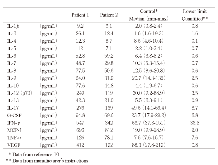
For serum cytokine measurement, blood samples were obtained from both patients at admission. Serum samples were frozen and stored at -80℃ until measurement of cytokine levels. All samples were measured on the same plate at the same time using the Bio-Plex Pro multiple cytokine measurement kit (Bio-Rad Laboratories Inc., Hercules, CA, USA) following the manufacturer’s instructions. Cytokines that were measured included IL-1β, IL-2, IL-4, IL-5, IL-6, IL-7, IL-8, IL-9, IL-10, IL-12 (p70) , IL-13, IL-17, granulocyte colony-stimulating factor, interferon (IFN) -γ, monocyte chemoattractant protein (MCP) -1, tumor necrosis factor (TNF) -α, and vascular endothelial growth factor [10]. Previously, we used this measurement system to evaluate serum samples of healthy children (median age 12 [range 4-21] months) , and we used the results as normal controls for infants and young children[10]
Results of cytokine assay are shown in Table 2. Serum levels for 13 of 17 cytokines in both patients were higher than the maximum level in normal controls. Specifically, levels of MCP-1 were more than 20 times higher than the maximum level in controls (695 and 812 vs 28.9 pg/mL) . In addition, both inflammatory cytokines (IL-6: 52.8 and 59.2 vs 8.2 pg/mL, TNF-α: 126 and 78.1 vs 16.7 pg/mL) and anti-inflammatory cytokines (IL-10: 77.6 and 44.8 vs 6.7 pg/mL) were remarkably increased in both patients compared with controls (Table 2)
Table. 1
Patient characteristics
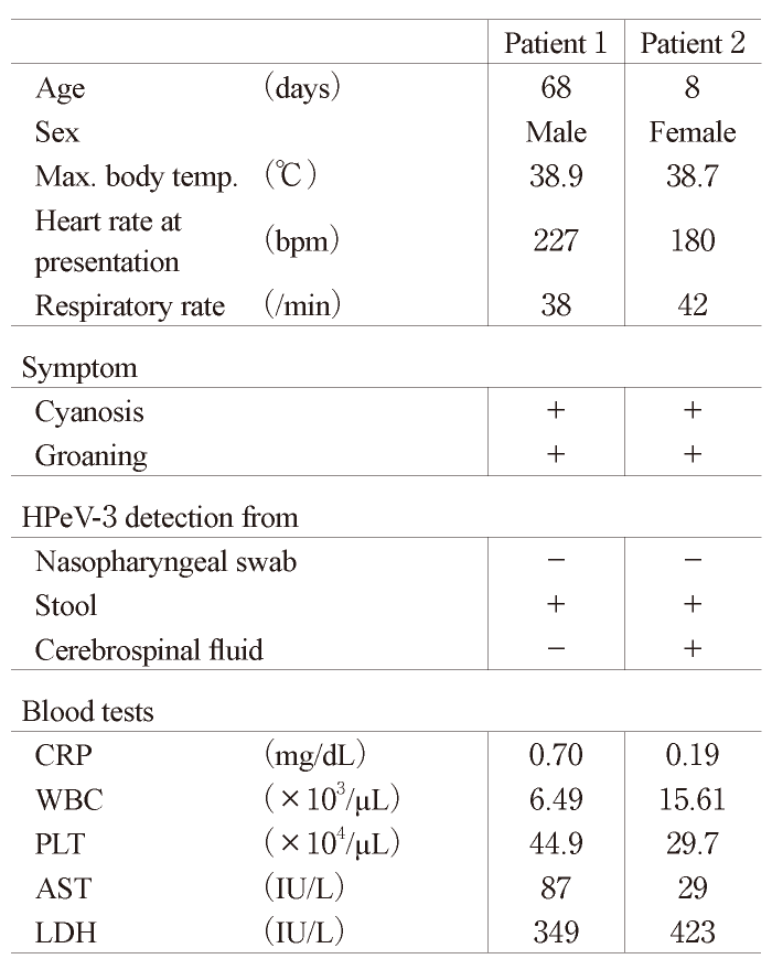
Table. 2
Cytokine concentration in the serum of the 2 infants with HPeV3 infection
Yuzurihara et al. [6]reported that the characteristic laboratory findings of patients with HPeV-3 infection were elevated levels of serum aspartate aminotransferase (AST) , lactate dehydrogenase, fibrin degradation, D-dimer, and ferritin, suggesting that organ damage was a result of hypercytokinemia-induced systemic cellular activation. Aviner et al. [7]described an infant with hemophagocytic lymphohistiocytosis associated with HPeV-3 meningitis. Serum levels of AST, D-dimer, fibrinogen, and ferritin were extremely high[7]. Hyperferritinemia was also reported in 6 patients with HPeV-3 infection complicated by enterovirus infection [8]. Results in our cases show direct evidence of hypercytokinemia, consistent with the findings of these previous reports.
We previously reported serum cytokine levels in patients with respiratory syncytial virus (RSV) lower respiratory tract infection, a common viral respiratory infection in infants[10]. RSV infection is also a common febrile illness in children, but IL-1β, IL-5, IL-6, IL-7, IL-12 (P70) , IL-13, IL-17, IFN-γ, MCP-1, and TNF-α levels in the 2 infants were also found to be high compared with levels in patients with RSV infection[10], suggesting cytokine storm with elevated levels of various inflammatory cytokines in systemic circulation. Overall, our data suggest that several inflammatory pathways can contribute to severe sepsislike conditions in neonates and infants.
Recently, the serum cytokine profile of HPeV-3 infection was reported for the first time. Shimizu et al. [11]measured serum levels of cytokines-IL-6, neopterin, IL-18, solube forms of tumor necrosis factor receptor types I (sTNFR-I) and II (sTNFR-II) - in 12 patients with sepsis-like symptoms at the onset, peak, and recovery phases. They concluded that proinflammatory cytokines, in particular, IFN-γ, TNF-α, and IL-18, are closely related to the development of HPeV3-induced sepsis-like syndrome. In both our cases, comprehensive evaluation of the laboratory data obtained from samples taken at a single timepoint in the early stage of the disease detected 17 kinds of inflammatory cytokines. Our data for IL-6 were similar to Shimizu et al.’s results and were considerably elevated in the acute phase. IL-6 is secreted by T cells and macrophages, and is a key molecule for humoral immunity. IL-6 is upregulated in many infectious and auto-immune inflammatory diseases, and is an indicator of disease severity[12]. At the same time, IL-10 levels, which have inhibitory roles in the immune response, were elevated. These anti-inflammatory cytokines are secreted rapidly in response to severe inflammation[13]. Elevated levels of IFN-γ and TNF-α in both our cases are also consistent with Shimizu et al.’s report [11].
The novel findings presented here include the remarkably elevated MCP-1 levels in HPeV-3 infection. MCP-1 is associated with eosinophil activation and migration. Elevated levels of this chemokine can result from activation in the initial phase in the immune system of neonates [14]
Some limitations might restrict the applicability of our results. First, we investigated only 2 patients. This is a pilot case study and a larger number of patients are needed to confirm these results and draw conclusions on the association between serum cytokine profiles and disease phenotypes. Second, Patient 1 was diagnosed with HPeV-3 infection by PCR assay from a stool sample. We did not perform PCR using blood samples, and the result was negative from the CSF sample. The diagnosis of HPeV-3 infection in this case was based on the characteristic phenotype and clinical course; however, the diagnostic mode could be insufficient. Infants with severe enterovirus infection may present similarity[15]. Therefore, additional investigations that compare the pathophysiology of HPeV-3 and enterovirus infection are necessary to confirm the clinical manifestation characteristic of HPeV-3 infection in neonates and infants.
In conclusion, our findings in these 2 cases suggest that severe sepsis-like conditions in HPeV-3 infections are caused by cytokine storm.
K.S. carried out patient care and wrote the manuscript. A.O. performed the diagnostic assay (PCR) for patients. J.T. critically reviewed the paper. H.H. measured cytokine levels and supervised the writing of the manuscript. All authors approved the final manuscript.
This study was supported by a grant from the Chiba Serum Institute Memorial Fund (H.H.)
All authors declare that there is no conflict of interest related to this study.
This study was originally planned as a clinical study and was approved by the Ethics Committee of Tokyo Women’s Medical University (#1492) . Written informed consent including that for publishing clinical information was obtained from parents or guardians.
All relevant data are available in the manuscript.
Address correspondence to Dr. Hiromichi Hamada.
Department of Pediatrics, Yachiyo Medical Center, Tokyo
Women’s Medical University, 477-96, Owada shinden, Yachiyo,
Chiba, 276-0046, Japan
Phone: +81-47-450-6000. Fax: +81-47-458-7047.
E-mail:hamada.hiromichi@twmu.ac.jp
