


Chiba Medical J. 97E:25-30, 2021
doi:10.20776/S03035476-97E-3-P25
〔 Original Short Communication 〕
Atsuro Yamazaki1) , Yusuke Matsuura1) , Kazuki Kuniyoshi1)
Takane Suzuki2) , Tomoyo Akasaka1) , Ei Ozone1)
Yoshiyuki Matsuyama1) , Michiaki Mukai1) , Takahiro Yamazaki1)
Takeru Ohara1) , Takahisa Sasho1) , and Seiji Ohtori1)
1) Department of Orthopaedic Surgery, Graduate School of Medicine, Chiba University, Chiba 260-8670.
2) Department of Bioenvironmental Medicine, Graduate School of Medicine, Chiba University, Chiba 260-8670.
(Received November 6, 2019, Accepted April 12, 2021, Published June 10, 2021.)
It is important to know the relationship between flexor tendon traction force and applied finger flexion force generated during the rehabilitation of trigger finger. However, there has been no report on this relationship using the Jamar dynamometer in clinical practice and cadaveric study. Therefore, the purposes of this cadaveric study were to measure the value of flexion force when pulling a tendon via the method used in clinical practice and to investigate the relationship between the traction force of the flexor tendon and the flexion force of the finger output based on the Jamar dynamometer. In this study, each finger of a fresh-frozen cadaver was pulled, and the finger flexion force was measured with the Jamar Plus+ Digital Hand Dynamometer (Performance Health, Chicago, IL, USA) . There was a strong first-order correlation between the flexor tendon traction force and the finger flexion force, and the value of the finger flexion force[N] divided by the flexor tendon traction force[N] was 0.195-0.321. Under the same flexor tendon traction force, the exerted finger flexion force was in the following order: middle finger, index finger, ring finger, and little finger (maximum to minimum) . It is important to consider these findings when performing rehabilitation of trigger finger.
Tendon force, finger flexion force, Jamar dynamometer, tendon traction force, cadaver
It is important to know how much force is applied to each tendon and joint during trigger finger’s rehabilitation, especially during A1 pulley stretching that has recently been implemented as a conservative treatment for trigger finger[1].
A1 pulley stretching is a clinically beneficial physical exercise. It requires proximal interphalangeal and metacarpophalangeal joint flexion with resistance achieved by fully grasping a block. The combined force of the tendon traction force and the reaction force pulls the A1 pulley to the palmar side and enlarges the A1 pulley lumen (Fig. 1) . Our previous study with freshfrozen cadavers has already verified the process leading to the enlargement of the A1 pulley lumen by this stretch[2]. In that study, traction force of up to 150 N was applied to the flexor tendon, and the expansion of A1 pulley lumen cross-sectional area was observed with the increase in traction force[2]. However, it has not been verified whether the value of flexion force produced by the traction force of up to 150 N, such as the A1 pulley stretching, is sufficient or not during the rehabilitation of trigger finger.
Verification of this issue requires knowledge of the relationship between the flexor tendon traction force and the finger flexion force generated. In this respect, there have been reports on the evaluation of this relationship using mathematical finger models[3], the flexor tendon under open carpal tunnel release[4], and flexor tendons of cadavers[5]. However, there is no report on this relationship using the Jamar dynamometer in clinical practice. Therefore, the purposes of this study were to measure the value of flexion force when pulling a tendon via the method used in clinical practice, and to examine the relationship between the tendon traction force and the finger flexion force.
We included 14 tendons from one fresh-frozen cadaver of a 100-year-old woman; these were two flexor digitorum profundus (FDP) and two flexor digitorum superficialis (FDS) tendons from the index, middle, and ring fingers, and one FDP and one FDS tendon from the little finger. All specimens were free from signs of trauma, deformity, and prior surgery. The cadaver had been kept at -20 ℃ and had not been frozen repeatedly. The cadaver was thawed just before the experiment and the tendons were dissected from the middle of the upper arm.
Using 0.118-inch-diameter Kirschner wires, we fixed the forearm in a neutral position with respect to the jig, and the wrist in a neutral position with 90° flexion. A dynamometer (Jamar Plus+ Digital Hand Dynamometer; Performance Health, Chicago, IL, USA) was placed on the palm (Fig. 2) . We made a skin incision on the anterior forearm and identified the FDP and FDS tendons. The FDP and FDS tendons were cut at the junction of the muscles and tendons. Traction tests were performed on the tendons using a nylon wire (diameter: 0.660 mm) . One end of the wire was attached to a universal testing machine (Autograph AG- 20kN X Plus; Shimadzu, Kyoto, Japan) . Traction was applied, and measurements were taken after the other end of the wire was sutured to the distal end of an FDS tendon. The right or left FDS tendon was each pulled toward the proximal end at a speed of 1.0 mm/s through the stiff string until a traction force of 150 N was recorded at 100 Hz or until the tendon ruptured. The dynamometer recorded the values of the FDS tendon traction force and the finger flexion force. This process was repeated in the FDP tendon. The average value of linear approximation equations was calculated for each tendon. No distinction was made between the FDS and FDP tendons’ average values in each finger, and both the values were designated as that of the particular finger. Hence, the average value of the linear approximation equations for each FDP and FDS tendon of the same finger was calculated as the average value for that finger. The model of ordinary least squares was used for the calculation of the approximate expression.
A strong first-order correlation was observed between the flexor tendon traction force and the finger flexion force recorded by the Jamar Plus+ Digital Hand Dynamometer for each tendon and for each finger (Fig. 3) . The R2 values were as follows: index finger, 0.999; middle finger, 0.989; ring finger, 0.993; and little finger, 0.964. The average value of the dynamometer [N] divided by the value of flexor tendon traction force [N] was as follows: index finger, 0.275; middle finger, 0.321; ring finger, 0.264; and little finger, 0.195. Under the same flexor tendon traction force, the maximum to minimum exerted finger flexion force was in the order middle finger, index finger, ring finger, and little finger.
Dennerlein et al.[6]examined the relationship between the FDS tendon traction force and the fingertip force during carpal tunnel release and reported that flexor tendon traction force increased proportionally with an increase in the intentional finger flexion force. Intrinsic muscles and extensor tendons may also be involved in finger flexion. However, from a biomechanical perspective, the extrinsic finger flexors, FDP and FDS, comprise the main sources of power for finger flexion in grasping-type motions, especially the power grip. Freivalds[7], Armstrong and Chaffin[8], and Cailliet[9]reported that the effects of intrinsic muscles and extensors on finger flexion can be disregarded because these muscles are typically in a relaxed state during the normal range of motion for a power grip. Based on the present experiment and these previous reports, the finger flexion force calculated during rehabilitation is proportional to the flexor tendon traction force. Dennerlein et al.[6]reported that the average ratio of the finger flexion force to the flexor tendon traction force ranged from 0.17 to 0.59 in nine subjects. Thus, the finger flexion force is in proportion to the flexor tendon traction force, and the result of the present study, in which the flexor tendon traction force was higher than the finger flexion force exerted, is similar to that in the previous study[6]. In addition, it was newly shown that a linear correlation could be established between the numerical value obtained by the Jamar Plus+ Digital Hand Dynamometer during the rehabilitation and the traction force of the flexor digitorum tendon.
In this study, a force of up to 150 N was applied to pull the flexor tendons. In previous research[3,6], it has been argued that the fingertip force is approximately one-third the tendon force. If 150 N of traction force is applied to all eight (for each finger, one FDP and one FDS separated from each other) flexor tendons on the index, middle, ring, and little fingers, the total traction force of all four fingers will be 1200 N. If the relationship between the above finger flexion force and flexor tendon traction force is used, the actual grip force is expected to be 400 N (40.8 kgf) . A grip force of 400 N is within the normal range in a typical adult observed in clinical practice[10]; hence, a traction force of as low as 150 N in each tendon is sufficient for the rehabilitation of trigger finger. Flexor tendon traction force at 0-150 N tested in our former study[2] was sufficient to produce a finger flexion force when performing stretching during rehabilitation. Since the expansion of the cross-sectional area of the A1 pulley lumen has been accompanied by an increase in tendon traction for a traction force of up to 150 N, it is expected that improvement can be achieved in individuals with stenosis at the A1 pulley level by exercising with a higher finger flexion force.
Our study showed that the bending force exhibited tended to be in the order middle, index, ring, and little fingers (maximum to minimum) under the same traction force. It is expected that the flexion force exerted under the same traction will increase in the order little, ring, index, and middle fingers, at least in the position that holds the Jamar-type dynamometers, as in our study. In addition, when performing an exercise that exerts finger flexion, such as A1 pulley stretching, if the same finger flexion force is used with each finger, the flexor tendon traction force of each finger conversely increases in the order little, ring, index, and middle fingers. This factor should be considered during rehabilitation and medical examinations.
This study has several limitations. First, the number of samples was insufficient to allow statistical comparison. However, a strong correlation was shown for all fingers and tendons, and a sufficient number of samples was used to confirm this relationship. Second and especially, statistically independent analysis of the FDS and FDP in each finger waits for further investigation. However, it was clear that the finger flexion force was proportional to the traction force for both the FDS and FDP. The ratio of the finger flexion force to the flexor tendon traction force is likely different between the FDS and FDP. Third, although we revealed the relationship between the finger flexion force, which arises as a result of applying traction to the tendon, and the traction force, it is unclear how much the intrinsic muscle, extensor tendon, and other soft tissues are involved when the finger applies a certain amount of flexion force. Lastly, it cannot be accurately determined how much traction force is actually applied to the flexor tendon. Since we know the approximate relationship between the finger flexion force in isometric finger flexion and the flexor tendon traction force, it is useful as a decisive factor for exercise intensity during rehabilitation.
We measured the value of finger flexion force when pulling a flexor tendon using the clinically followed method of using the Jamar dynamometer. A strong first-order correlation was observed between the flexor tendon traction force and the finger flexion force. Under the same flexor tendon traction force, the maximum to minimum finger flexion force is in the following order: middle, index, ring, and little fingers. It is important to consider these findings when performing rehabilitation.
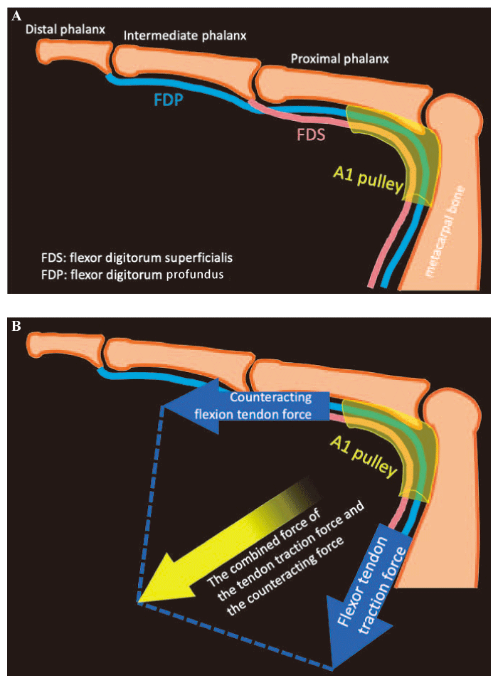
Fig. 1. The mechanism of A1 pulley stretching. A. The relationship between bones, flexor digitorum superficialis (FDS) , flexor digitorum profundus (FDP) , and A1 pulley. B. The combined force of the flexor tendon traction force and the counteracting force pulls the A1 pulley to the palmar aspect and enlarges the A1 pulley lumen.
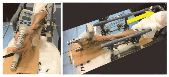
Fig. 2. Jamar Plus+ Digital Grip Hand Dynamometer and upper extremity position. The FDP and FDS tendons were pulled proximally (arrow) at a speed of 1.0 mm/s through the stiff string until 150 N of traction force was recorded at 100 Hz (A. Front view, B. Side view) .
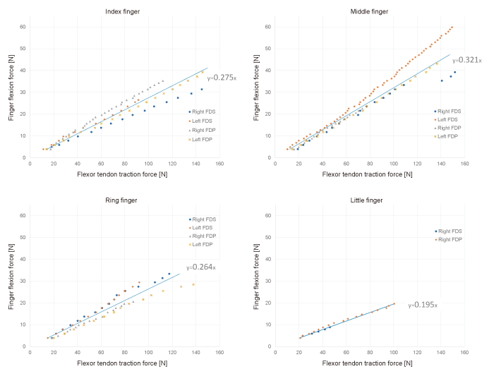
Fig. 3. The average of linear approximation equations of the relationship between the flexor traction force and the finger flexion force for each tendon. A strong first-order correlation was observed between the flexor traction force and the finger flexion force recorded by the Jamar Plus+ Digital Hand Dynamometer for each tendon and for all fingers (maximum R2 = 0.999, minimum R2 = 0.964) . The left FDS and FDP of the little finger were too small to be investigated.
A. Y., Y. Matsuu., K. K., T. Su., T. A., E. O., Y Matsuy., M. M., T. Y., O. T., T. Sa., and S. O. substantially contributed to the research design and data analysis. All authors have read and approved the final submitted manuscript.
The authors received grant funding from the Japan Society for Surgery of the Hand.
S.O. is a member of the Editorial Board of the Chiba Medical Journal. The other authors declare that they have no conflicts of interest, either financial or nonfinancial, with the contents of this article.
The research protocol was in compliance with the Helsinki Declaration; it was approved by the Research Ethics Committee of Graduate School of Medicine, Chiba University (Authorization number: #2815) and registered with the University Hospital Medical Information Network. Written informed consent was obtained from the donor before death.
The data that support the findings of this study are available from the corresponding author, upon reasonable request.
We are deeply grateful to the Shiragikukai Association for anatomical donation and members of the Department of Bioenvironmental Medicine, Graduate School of Medicine, Chiba University.
Address correspondence to Dr. Atsuro Yamazaki.
Department of Orthopaedic Surgery, Graduate School of Medicine,
Chiba University, 1-8-1 Inohana, Chuou-Ku, Chiba 260-8670, Japan
Phone: +81-43-226-2117. Fax: +81-43-226-2116.
E-mail:atsuroh1984@gmail.com
