


Chiba Medical J. 97E:75-83, 2021
doi:10.20776/S03035476-97E-6-P75
〔 Chiba Medical Society Award Review 〕
Takayuki Baba
Department of Ophthalmology and Visual Science, Chiba University Graduate School of Medicine, Chiba 260-8670.
(Received September 11, 2021, Accepted September 27, 2021, Published December 10, 2021.)
The population with high myopia is growing worldwide, especially in East Asia. In eyes with pathologic myopia, the axial length is elongated and the eyeball’s shape resembles a rugby ball. The posterior pole of the eye has an outpouching structure, and this unique shape is called a posterior staphyloma. The posterior staphyloma affects the pathogenesis of myopic tractional maculopathy (MTM) . MTM is caused by traction on the macula and develops retinal schisis, macular hole, and retinal detachment due to a macular hole. The prevention of advanced stages of MTM, such as retinal detachment, is essential. As the tractional force on the retina is mainly caused by posterior staphyloma and posterior vitreous cortex/internal limiting membrane (ILM) , two approaches, scleral surgery and vitrectomy, are currently used. Although there have been many surgical options for treating eyes with MTM, the standard approach for this pathology is yet to be established. Less invasive surgery is ideal because myopic schisis is an early stage of MTM in patients with relatively good vision, and secondary macular hole and visual decline are sometimes observed after surgery for myopic schisis. We used scleral imbrication and achieved significant visual recovery with less chance of developing a secondary macular hole.
myopic traction maculopathy, retinal detachment, schisis, high myopia, scleral imbrication
In highly myopic eyes, the length of the eyeballs is longer than that of the emmetropic eyes. The axial length (AL) of eyes with high myopia is more than 26.0 or 26.5 mm, which is longer than that of emmetropic eyes by two millimeters or more (Fig. 1A). The population with high myopia is growing worldwide, especially in East Asia. Approximately 22.9% of the global population had myopia greater than -0.5 diopter (D) in 2000, and half of the global population (49.8%) is estimated to be myopic by 2050[1]. The increasing trend is similar in Japan; the prevalence of myopia greater than -0.5 D was 50.0%, and -6.0 D was 7.9% in a cohort study conducted between 2013-2016. The population of myopia (> -0.5 D) has significantlyincreased from 41.8% in 2000[2]and 37.7% in 2005[3]. Eyes with low-grade myopia have a morenegligible effect on corrected visual acuity. On the other hand, high myopia (> -6.0 D) causes pathologicchanges, including myopic choroidal neovascularization, myopic tractional maculopathy (MTM) , chorioretinal atrophy, and myopic neuropathy[4]. The pathologic alterations result in severe vision impairment, which is called pathologic myopia. In this review, I focus on MTM, a potentially blinding disease, and discuss the strategy for the treatment of this refractory disease.
In eyes with pathologic myopia, the AL is elongated, and the eyeball resembles a rugby ball. The posterior part of the eyeball is significantly elongated compared to the anterior segment. The posterior pole of the eye has an outpouching structure, and this unique shape is called a posterior staphyloma. The size and kurtosis of the posterior staphyloma vary among the eyes[5]. Some eyes have multiple protrusions in the posterior staphyloma.
The posterior staphyloma affects the pathogenesis of MTM. When the axial elongation occurs, the posterior sclera expands posteriorly and forms the posterior staphyloma. However, the inner part of the eyewall, which consists of the choroid and retina, cannot follow the extreme expansion and is left behind at various degrees. This causes retinal traction anteriorly or toward the center of the eyeball (Fig. 2) . The internal limiting membrane (ILM) , the innermost layer of the retina, causes tangential traction to the retina and causes the pulling force anteriorly. The posterior vitreous cortex also causes anteroposterior and tangential traction on the retina, causing MTM. The retinal vessels, especiallythe retinal arteries, are relatively stiff and less resilient. Therefore, these vessels cannot follow the contour of the posterior staphyloma and cause traction on the retina. Collectively, these tractional forces work together on the retina and cause MTM.
MTM is a spectrum of diseases that begins with mild retinal thickening and myopic retinal schisis (Fig. 1B) . Myopic schisis was first described by close observation using a slit-lamp biomicroscope in 1958[6]. Because of the difficulty in observation, this pathology was not well accepted until optical coherence tomography (OCT) was available in the late 1990s. Myopic schisis is observed clearly using OCT[7]. The microstructure of myopic schisis is an increased retinal thickness with numerous pillar structures, which are considered Müller cells. The prevalence of myopic schisis has been reported to be 9-34% in patients with high myopia[8]. Myopic schisis is less symptomatic, and patients rarely notice visual impairment or metamorphopsia. However, one to 14% of eyes with myopic schisis develop a full-thickness macular hole, and 2-21% of cases develop retinal detachment associated with macular holes[9-11]. Patients with full-thickness macular holes suffer severe metamorphopsia and a decline in visual acuity of less than 0.1 (Fig. 1C) . Retinal detachment secondary to the macular hole is more aggressive and results in total retinal detachment, proliferative vitreoretinopathy, and blindness if the patients are not adequately treated (Fig. 1D) . The treatment for retinal detachment due to a macular hole remains challenging, even with advanced vitrectomy surgery[12]. Therefore, the prevention of advanced stages of MTM, such as retinal detachment, is essential. As the tractional force on the retina is mainly caused by the posterior staphyloma and posterior vitreous cortex/ILM, two approaches, scleral surgery and vitrectomy, are currently used.
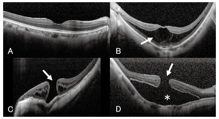
Fig. 1 Macular morphology in normal and myopic eyes (A) Optical coherence tomography shows a normal macular configuration in an eye with an axial length of 23.5 mm. Visual acuity is 1.0. (B) Myopic schisis is observed with a thickened retina, and multiple columnar structures (arrow) . Axial length is 26.8 mm and visual acuity is 0.4. (C) A macular hole is observed in an eye with an axial length of 33.4 mm (arrow) . Visual acuity is 0.1. (D) Retinal detachment (asterisk) associated with a macular hole (arrow) is observed. Axial length is 28.0 mm, and visual acuity is 0.03.
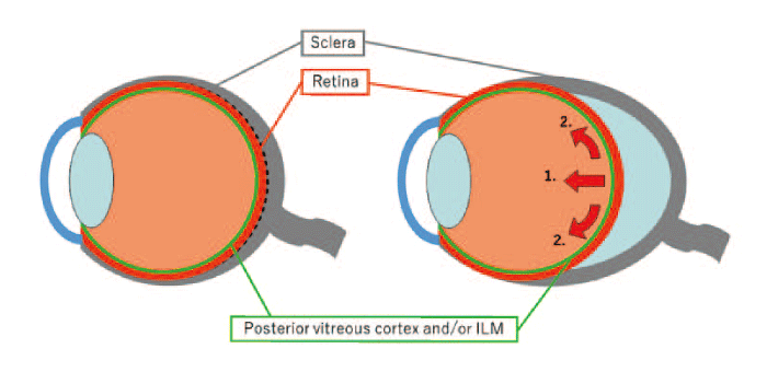
Fig. 2 Elongation of an eyeball in high myopia. The schematic shows an eye with a normal (left) and a longer (right) axial length. Anteroposterior traction by the vitreous (1.) and tangential traction by the posterior vitreous cortex and internal limiting membrane (ILM) (2.) cause the retinal detachment and schisis.
1 .Ando-plombe
Since MTM is frequently observed in eyes with posterior staphyloma, the abnormal expansion of the posterior eyeball suggests the vital role as a cause of MTM. It seems to be a clue for treating MTM to correct the shape of the posterior staphyloma. Scleral buckling is one approach to change the curvature of the sclera and is commonly used for treating rhegmatogenous retinal detachment (RRD) . In eyes with RRD, the retinal breaks are located at the peripheral retina and are easily sealed by scleral buckling because the surgical field is anterior and easy to access. However, the location of the posterior staphyloma is the deepest point of the eyeball, and surgical manipulation is challenging. To overcome this difficulty, the Ando plombe was designed. It is made of silicone rubber and a titanium-made stylet to maintain its shape[13]. Using this specially designed scleral buckle, surgeons can put pressure on the posterior staphyloma toward the inside (Fig. 3) . In 2006, we first used the Ando plombe to treat myopic schisis and reported the outcome[14]. We treated six eyes of five myopic schisis cases using this technique. We followed the visual acuity, fundus appearance, and structural changes observed using OCT every three months, postoperatively. As a result, retinal schisis was significantly reduced by six months after surgery. Four eyes (67%) showed a significant improvement in visual acuity by more than two lines, and the remaining eyes (33%) showed no significant change from the baseline. This favorable outcome was due to the protrusion created by the Ando plombe. One eye had a retinal hemorrhage with choroidal neovascularization, but visual acuity was retained during the follow-up period. We believed that this surgical technique was effective and could be an option for treating myopic retinal schisis. However, chorioretinal atrophy developed many years after the surgery, possibly because of the reduced choroidal circulation by the plombe. The plombe causes an abrupt change in the eyewall and possibly obstructs the blood flow of the choroid around it.
2 .Alterations of macular buckling
Other researchers used T-shaped solid silicone rubber [15], L-shaped silicone sponge using stainless steel for increased rigidity[16], and 3-armed silicone capsule[17]as a scleral buckle. The basic idea of these other forms of the scleral buckle was similar to ours, and the surgical outcome was favorable. However, it may be associated with the risk of chorioretinal atrophy in the long term. There is another type of scleral buckling that uses the human sclera instead of a silicone material. A scleral patch to reinforce the posterior staphyloma has also been reported[18]; a piece of sclera from the donor’s eyes was sutured onto the patient’s sclera. Eighty-three percent of the cases showed complete resolution of myopic schisis, and an improvement in visual acuity of more than 0.1 was obtained in 75% of the cases. The outcome seems favorable in the short term, but the long-term effects are unknown. The effect of the scleral patch is gradually lost because of the degradation of the transplanted sclera and loosening of the sutures.
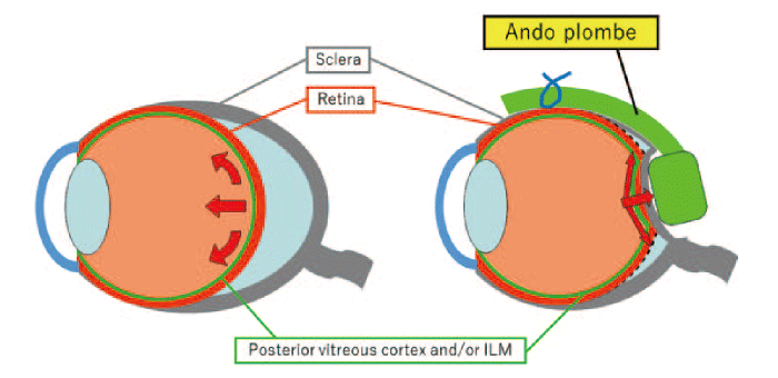
Fig. 3 Scleral buckling with the Ando plombe. The schematic shows scleral buckling surgery using the Ando plombe (right) . The plombe is sutured on the temporal sclera, which makes a protrusion on the macula. It should be noted that the direction of the traction force is reversed to reattach the retina. ILM: internal limiting membrane.
Retinal traction by the posterior vitreous cortex and an ILM is another critical factor in the development of MTM. Scleral buckling has the effect of inverting the tangential traction by changing the contour of the eyewall and exerting an outward force to resolve retinal schisis. In contrast, vitrectomy reduces tangential retinal traction by removing the posterior vitreous cortex and ILM (Fig. 4) . With the recent advancement in microincision systems, vitrectomy has become less invasive and widely spread worldwide.
1 . Vitrectomy with the removal of the ILM
Vitrectomy consisting of core vitrectomy, removal of the macular vitreous cortex and ILM in the posterior staphyloma, and gas tamponade with 30% SF6 was performed in nine eyes [19]. Eight of nine eyes showed the resolution of myopic schisis, and the visual acuity ranged from 0.4 to 0.6 at six months, postoperatively. One eye developed a full-thickness macular hole and showed a poor vision of 0.08. Ikuno et al. reported anatomical success in five of six eyes with significant visual recovery in all eyes [20]. Hirakata et al. reported visual improvement in nine of 16 eyes with myopic schisis and detachment after vitrectomy with posterior vitreous separation[21]; in their case series, two eyes developed macular hole retinal detachment. In summary, vitrectomy with removal of the ILM and posterior vitreous cortex effectively treats myopic schisis, but 11- 33% of cases develop macular holes with poor visual acuity after the initial surgeries[19,21,22]. A defect in the ellipsoid zone observed on preoperative OCT is thought to be predictive for the development of full-thickness macular holes after surgery[22]
2 . Fovea-sparing ILM peeling
The poor visual outcome in eyes that developed a full-thickness macular hole postoperatively resulted in a new technique, fovea-sparing internal limiting membrane peeling (FSIP) . This technique removes the ILM during vitrectomy, but the central part of the ILM is left as it is[23,24]. By leaving the ILM around the fovea, surgeons can avoid damaging the very thin part of the inner retina and prevent the secondary macular hole. A full-thickness macular hole developed in 16.7% of eyes after complete removal of the ILM, and none of the 15 eyes were treated using FSIP. In the FSIP group, the resolution of the schisis was partial before 12 months postoperatively, and complete resolution of myopic schisis was observed afterward. Visual recovery was significant in the FSIP group (P=0.04) , but not in the complete peeling group. This technique is relatively simple for experienced retinal surgeons and is gaining popularity in the current era of microincision vitrectomy surgery.
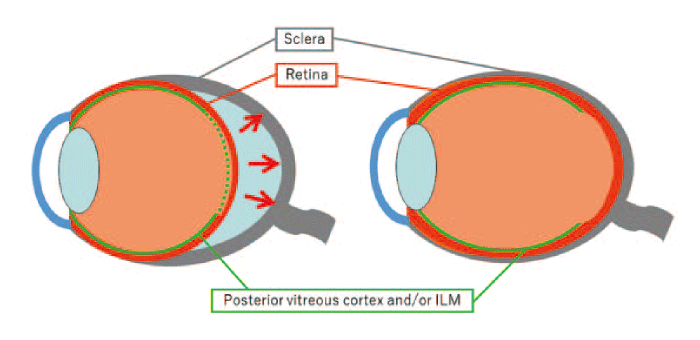
Fig. 4 Vitrectomy with the removal of the membrane. The posterior vitreous cortex and the internal limiting membrane (ILM) are removed (dotted line, left) . After this procedure, the retina softens and expands posteriorly (arrows) . Finally, the retina is attached to the eyewall (right) .
Scleral imbrication
Although vitrectomy using FSIP is a relatively safe approach for myopic schisis, some cases develop microscotomas[25]and macular hole[26]after surgery. These vision-threatening complications should be prevented, especially in cases with monovision (one eye has no vision due to disease or trauma) . For this purpose, we used another approach with the expectation of reducing complications such as microscotoma and macular hole; scleral imbrication, which involves the infolding of the sclera (Fig. 5) [27]. During the surgery, mattress sutures with 6 mm intervals were placed at the temporal quadrants of the sclera (Fig. 6A) ; 4-0 polyester ophthalmic sutures were used. Core vitrectomy was performed using a three-port system and a 25- or 27-gauge microincision vitrectomy system. After removal of the vitreous core, triamcinolone acetonide was used to visualize the vitreous cortex. In most cases, the remnants of the thin posterior vitreous cortex attach to the retinal surface. This thin membrane was removed using a silicone-tip needle or a diamonddusted membrane scraper to prevent damage to the retinal surface (Fig. 6B) . In some cases, we used microtipped forceps on the firmly adhered membrane. The intraocular pressure was then decreased to 5 mm Hg to make the eyeball soft. The mattress sutures were then tightened to infold the sclera (Fig. 6C) . The protrusion of the fundus was observed in the temporal area if imbrication was successfully performed (Fig. 6D) . This whole procedure could be performed under local anesthesia either by retrobulbar or sub-Tenon’s capsule injection of 0.5% levobupivacaine. We did not perform phacoemulsification and implantation of the intraocular lens if the cataract was not severe enough to disturb fundus visibility during surgery.
We used scleral imbrication in eight eyes of eight patients. Six eyes had retinal detachment and schisis, and the other two eyes had schisis only. The average patient age was 67.1 years (61-76 years) , and all patients were female. We examined the best-corrected visual acuity (BCVA) , AL, central foveal thickness, and curvature of the eyewall measured using OCT, preoperatively, and at 3, 6, and 12 months, postoperatively. BCVA was 0.71 ± 0.25 logMAR units (decimal acuity: 0.2) at the baseline, and 0.58 ± 0.27 logMAR units (decimal acuity: 0.25) at 3 months, 0.62 ± 0.33 logMAR units (decimal acuity: 0.25) at 6 months, and 0.36 ± 0.19 logMAR units (decimal acuity: 0.4) at 12 months, postoperatively (P=0.028, Fig. 7A) . Central foveal thickness decreased from 540 ± 171 μm at the baseline to 310 ± 96 μm at 3 months (P=0.012) , 206 ± 131 μm at 6 months (P=0.012) , and 170 ± 85 μm at 12 months (P=0.012, Fig. 7B) . AL shortened from 29.5 ± 1.7 mm to 27.4 ± 2.3 mm at 1 month (P= 0.012) , to 28.1 ± 2.2 mm at 6 months (P=0.012) , and to 28.5 ± 2.5 mm at 12 months (P=0.012, Fig. 7C) . The curvature of the eyewall is represented by a coefficient a in a second-order polynomial equation, and a large a represents the greater steepness of the curve.
y = ax2 + bx + c
The average coefficient a was 8.2 ± 1.2 x 10⁻⁴ at the baseline, 7.3 ± 1.7 x 10⁻⁴ at 6 months, and 7.4 ± 1.6 x 10⁻⁴ at 12 months (P=0.036 at 6 months and P=0.049 at 12 months, Fig. 7D) shows flattening of the posterior staphyloma after the surgery. Retinal detachment was completely resolved in five of six eyes (83%) by 12 months, and the remaining eye showed a decrease in height. No complications of secondary macular hole or RRD were observed after the surgery.
Scleral imbrication showed significant improvement in visual acuity without any severe complications. This safe technique is particularly beneficial for cases with monovision. The effect of scleral imbrication seems to be exerted by shortening the eyeball and flattening the posterior staphyloma (Fig. 5) . As MTM is caused by the elongation of the eyeball and deformity of the posterior part of the eyeball, scleral imbrication is a reasonable maneuver. The effect lasts for three or more years after the initial surgery[28]. The negative aspects of this surgery are surgery-induced astigmatism and diplopia. Astigmatism is caused by changes in the shape of the cornea and can be corrected using a contact lens or cataract surgery with implantation of a toric intraocular lens. Diplopia is likely noticed if the patient looks at the distance. The temporal rectus is weakened by scleral imbrication and causes diplopia. The use of spectacles with a prism lens or muscle surgery, such as plication of the temporal rectus, is effective. Diplopia never occurs in a patient with monovision, and it is another good reason for a better indication of this surgery for a patient with monovision with MTM.
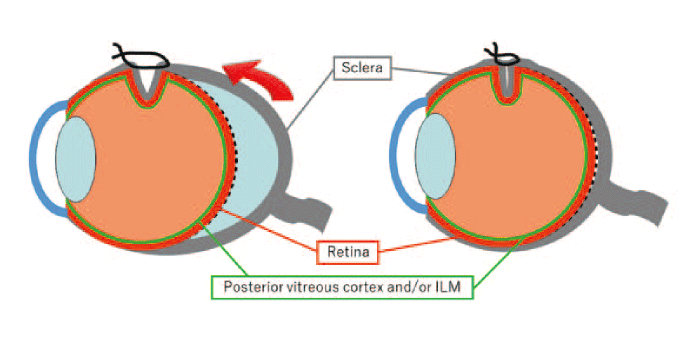
Fig. 5 Scleral imbrication. When the mattress sutures placed on the temporal sclera are tied up, the posterior part of the sclera is shortened toward the temporal area (arrow, left) . The shape of the eyeball is corrected, and the retinal schisis and detachment are resolved (right) . ILM: internal limiting membrane.
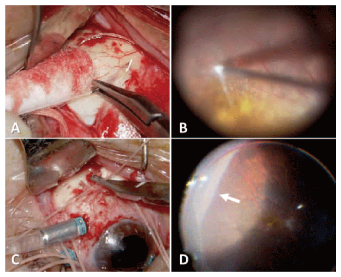
Fig. 6 Surgery of scleral imbrication. (A) Mattress sutures with 6 mm intervals are placed on the superior and inferior temporal sclera. (B) The posterior vitreous cortex is gently removed but the internal limiting membrane (ILM) is not removed. (C) The sutures are tightened with decreased intraocular pressure and the sclera is infolded. (D) Protrusion by scleral imbrication is observed at the temporal area (arrow) .
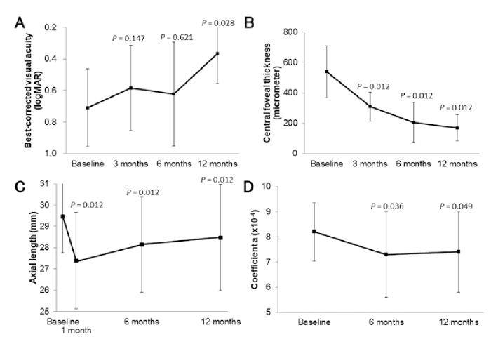
Fig. 7 Outcome after scleral imbrication. (A) The average best-corrected visual acuity is not improved significantly at 3 and 6 months postoperatively (P=0.147 and P=0.621, Wilcoxon test) . At 12 months postoperatively, the visual acuity is significantly better than that at the baseline (P=0.028) . (B) Central foveal thickness is significantly reduced at 3, 6, and 12 months postoperatively (P=0.012, 0.012, and 0.012, respectively) . (C) The average axial length is significantly shorter at 1 month after the surgery, and then gradually recovers to the original length. However, the axial length is still shorter than that of the baseline at 6 months (P=0.012) and 12 months (P=0.012) . (D) The steepness of the curve of the eyewall is represented by the coefficient a in a second-order polynomial equation, ax2 + bx + c. After the scleral imbrication, the steepness of the posterior eyewall slightly decreased (P=0.036 at 6 months and P=0.049 at 12 months) .
MTM is a devastating condition observed in patients with high myopia. The pathology is potentially progressive and requires intervention before progression to the unretrievable stage. Therefore, surgery for myopic retinal schisis is essential. Less invasive surgery is ideal because secondary macular holes and visual impairment are sometimes observed after surgeries for myopic schisis. Scleral imbrication has shown a significant recovery of vision with a lower chance of developing secondary macular holes. The importance of scleral imbrication surgery should be confirmed with a large number of cases, especially in those with monovision, in the future.
None.
The author declares no conflict of interest associated with this manuscript.
Not applicable.
Not applicable.
The author expresses gratitude to Prof. Shuichi Yamamoto for critically reading the manuscript.
Address correspondence to Dr. Takayuki Baba.
Department of Ophthalmology and Visual Science, Chiba University
Graduate School of Medicine, 1-8-1 Inohana, Chuoku,
Chiba 260-8670, Japan.
Phone: +81-43-226-2124. Fax: + 81-43-224-4162.
E-mail:t.baba.oph@faculty.chiba-u.jp
