


Chiba Medical J. 98E:29-37, 2022
doi:10.20776/S03035476-98E-4-P29
〔 Original Article 〕
Masaya Mizutani1), Yawara Eguchi1,2), Sumihisa Orita1,3)
Kazuhide Inage1), Yasuhiro Shiga1), Satoshi Maki1)
Junichi Nakamura1), Shigeo Hagiwara1), Yasuchika Aoki4)
Masahiro Inoue4), Masao Koda5), Hiroshi Takahashi5)
Tsutomu Akazawa6), and Seiji Ohtori1)
1) Department of Orthopaedic Surgery, Graduate School of Medicine, Chiba University, Chiba 260-8670.
2) Department of Orthopaedic Surgery, Shimoshizu National Hospital, Yotsukaido 284-0003.
3) Center for Frontier Medical Engineering, Chiba University, Chiba 263-8522.
4) Department of Orthopaedic Surgery, Eastern Chiba Medical Center, Togane 283-8686.
5) Department of Orthopedic Surgery, University of Tsukuba, Tsukuba 305-8575.
6) Department of Orthopaedic Surgery, St. Marianna University School of Medicine, Kawasaki 216-8511.
(Received March 24, 2022, Accepted June 10, 2022, Published August 10, 2022.)
【Purpose】The incidence of Tandem Spinal Stenosis (TSS) is increasing rapidly with population aging, though its pathophysiology and pathogenesis are not clear. There is no consensus whether a one-stage or two-stage procedure is better. We compared preoperative skeletal muscle mass, clinical symptoms, and surgical results between TSS patients who underwent simultaneous cervical and lumbar decompression and lumbar spinal stenosis (LSS) patients who underwent lumbar decompression. The characteristics of the TSS patients and the surgical outcomes of one-stage surgery were evaluated.
【Methods】There were 118 patients (mean age 66.3 years, 71 men) who received surgical treatment. Of these, 9 patients had TSS (mean age 74.3 years, 4 men) and 109 had LSS (mean age 65.7 years, 67 men). A bioelectric impedance analyzer (BIA) was used to measure systemic skeletal muscle mass and phase angle, an index of cell membrane aging. The visual analog scale (VAS) score for low back pain (LBP) and leg pain and leg numbness, the Japanese Orthopaedic Association scoring system (JOA score), the Roland-Morris Disability Questionnaire (RDQ), the Japanese Orthopaedic Association Back Pain Evaluation Questionnaire (JOABPEQ), and the Oswestry Disability Index (ODI) were used to evaluate clinical symptoms. Preoperative skeletal muscle mass, phase angle, clinical symptoms, and surgical results were compared between the groups.
【Results】Operative time was significantly longer in the TSS group than in the LSS group (p < 0.05), but the amount of bleeding was not significantly different between the groups. The height, trunk muscle mass, and phase angle of the trunk and both lower limbs were significantly lower in the TSS group than in the LSS group (p < 0.05). JOA scores were significantly lower in the TSS group (p < 0.05), and 88% of TSS patients had difficulty in climbing stairs, significantly more than in the LSS group (28%, p < 0.05). Although clinical symptoms in both groups improved after surgery, there was no significant difference in the degree of clinical improvement (p > 0.05).
【Discussion】TSS was associated with decreased trunk muscle mass and decreased phase angle of the trunk and both lower limbs, suggesting the progression of trunk muscle atrophy due to decreased activity. In addition, about 90% of TSS patients had difficulty in climbing stairs. A one-stage minimally invasive operation was performed for TSS and the amount of intraoperative bleeding was equivalent to that of LSS alone. The one-stage approach seems to be effective.
Tandem Spinal Stenosis, Lumbar Spinal Stenosis, skeletal muscle, cell aging, disability
In our aging society, the number of patients with spinal stenosis is increasing annually. Spinal stenosis is often caused by age-related changes such as disc degeneration and osteophyte formation, which are often found in the highly mobile cervical and lumbar spine[1- 3]. The prevalence of cervical spondylotic myelopathy (CSM) is estimated to be 5% to 20%, and lumbar spinal stenosis (LSS) is estimated to be 8% to 11%[4,5]. However, it has been reported that a significant percentage of patients, including asymptomatic elderly patients, are observed to have stenosis on simple X-ray. Clinical symptoms differ depending on whether the stenosis site is outside or in the center of the spinal canal, or in the intervertebral foramen[6]. Tandem Spinal Stenosis (TSS) is observed in both the cervical and lumbar spine and is accompanied by varied symptoms.
TSS was first reported by Teng and Papatheodorou in 1964[7]. Dagi et al. defined TSS based on three clinical signs: (1) intermittent lower extremity claudication, (2) the mixed motor neuron sign of upper and lower limbs, and (3) progressive gait disturbance[8]. The prevalence is reported to be high in women, ranging from 7.6% to 60% of patients with spinal stenosis[7-14]. Kawaguchi et al. have reported an association between ossification of posterior longitudinal ligament (OPLL) and TSS[9], but the causes and risk factors for TSS are not yet clear. Conservative treatment or surgical treatment is selected according to the symptoms, but there is still no consensus on whether surgical treatment should be a one-stage or a two-stage approach.
The purpose of this study was to compare preoperative skeletal muscle mass, clinical symptoms, and surgical results between TSS patients who underwent simultaneous cervical and lumbar decompression and LSS patients who underwent lumbar decompression only. In addition, the characteristics of TSS patients and the effectiveness of onestage surgery were examined.
Participants
Written informed consent was obtained from all participants before the study began. The study protocol was approved by the ethics review committee (IRB approval no., H26’-4).
From November 2017 through January 2021, 118 patients who visited our outpatient department of orthopedics, were diagnosed as LSS or TSS, received surgical treatment, and were followed up for six months or more were included in the study. Of the 118 patients (mean age 66.3 years, range 19-87 years, 71 men), 9 patients (mean age 74.3 years, range 62-85 years, 4 men) were diagnosed as TSS, and 109 patients (mean age 65.7 years, range 19-87 years, 67 men) were diagnosed as LSS.
In the TSS group, six patients had 3-level cervical decompression, two had 4-level decompression, and one had 5-level decompression. In the lumbar region, three had single-level decompression, four had 2-level decompression, and one had 3-level decompression.
In the LSS group, 76 patients had single-level lumbar decompression, 19 had 2-level decompression, 12 had 3-level decompression, and two had 4-level decompression.
Lumbar spinous process-splitting laminectomy (L-SPSL)[15]was performed for LSS. For TSS patients, myovascular preserving open-door laminoplasty with mini-plate fixation (MPLP)[16]was performed on the cervical spine and L-SPSL was performed on the lumbar spine. Patients were positioned prone with their heads secured using Mayfield clamps. A single spine surgeon (Y.E.) performed the decompression surgeries.
Patients who had previous spinal surgery and those with spinal tumor, infectious disease, or spinal trauma were excluded.
Analysis of skeletal muscle mass and phase angle
A multifrequency bioelectrical impedance analyzer (BIA; MC-780A, TANITA, Tokyo, Japan) was used according to the manufacturer’s guidelines. BIA is a noninvasive examination technique used for evaluating bone mass, fat mass, and fat-free mass by flowing weak currents with three different frequencies (5, 50, and 250 kHz) using 8 electrodes in total, two for each sole and grip in a standing barefoot position and determining the difference in electric resistance. The analysis time is less than 20 s. Limb and trunk muscle masses were determined directly from the value of lean mass provided by the device.
Appendicular skeletal muscle mass was calculated as the sum of skeletal muscle mass in the arms and legs, assuming that the mass of lean soft tissue is effectively equivalent to skeletal muscle mass. Appendicular skeletal mass index (SMI) was determined as the sum of arm and leg lean mass (kg) / ( height (m) )2.
Resistance (R) and reactance (Xc) measured at 50 kHz were used to calculate the phase angle using the following equation: Phase angle (degrees) = - arctangent (Xc / R) × (180 / π).
Clinical symptoms
The visual analog scale (VAS) score for low back pain (LBP), leg pain, and leg numbness, which ranges from 100 mm (severe pain) to 0 mm (no pain), the Japanese Orthopaedic Association (JOA; 0-29 points), and the Roland-Morris Disability Questionnaire (RDQ; 0-24 points) were used to assess clinical symptoms. The normal JOA score is 29 points, based on three subjective symptoms (9 points), three clinical indicators (6 points), and seven activities of daily living (14 points). The standard RDQ is zero points, with the total number of items checked ranging from 0 to 24.
The Japanese Orthopaedic Association Back Pain Evaluation Questionnaire (JOABPEQ) consists of 25 RDQ-based questions and the Short Form 36. (SF-36).
A symptom score of Q1-1 through Q4-1 and Q5-1 was defined as positive, and a symptom score of “2” or “3” was defined as negative. A score of Q4-2, Q4- 3, Q5-2 to Q5-7 was defined as positive if it was “1” or “2”, and a score of “3” to “5” was defined as negative. Answers to questions in five domains are used to construct scores: pain-related disorders, lumbar spine dysfunction, gait disturbance, social life dysfunction, and psychiatric disorders. Each domain’s score was calculated according to official rules and ranged from 0 to 100 points, with the score being proportionate to the patient’s clinical condition.
Statistical Analyses
In both groups, we investigated operative time, intraoperative bleeding volume, age, height, weight, BMI, body fat percentage, skeletal muscle mass, phase angle, and clinical symptoms. An unpaired t test was used to assess differences between groups for each variable and clinical symptom. For each JOABPEQ item, differences between groups were evaluated using the chi-squared test. All data are expressed as the mean ± standard deviation (SD). P < 0.05 was considered significant. Statistical analyses were performed using SAS for Windows (Ver. 9.4, SAS Institute Inc., Cary, NC, USA).
Operative time (min) was 159.3 ± 31.9 in the TSS group and 88.1 ± 40.7 in the LSS group (p < 0.001). The intraoperative bleeding volume (ml) was 103.4 ± 30.7 in the TSS group and 68.2 ± 55.0 in the LSS group. There was no significant difference (p = 0.076).
On the physical examination, both height (p = 0.028) and truncal muscle mass (p = 0.044) were significantly greater in the LSS group than in the TSS group. No other physical characteristics were statistically different (Table 1).
Phase angles of the left half body (p = 0.048), right lower limbs (p = 0.039), left lower body (p = 0.036), and both lower limbs (p = 0.042) were significantly more negative in the LSS group than in the TSS group. There was no significant difference between groups for the right or left hands (Table 1).
Preoperatively, VAS score for leg pain (p = 0.007) and JOA score (p = 0.016) were significantly higher in LSS than in TSS (Fig. 1). There were no significant differences between groups for the other VAS scores, RDQ score, or ODI score. There were no significant differences between the groups in any broad category of the JOABPEQ, including Pain-related disorders, Lumbar spine dysfunction, Gait disturbance, Social life disturbance, or Psychological disorders (Fig. 2). However, patients in the TSS group had significantly more difficulty climbing stairs than did those in the LSS group (Table 2).
The JOA score was significantly better (p = 0.028) and the VAS score for leg pain was significantly lower (p = 0.021) following surgery in the TSS group (Fig. 3). In the LSS group, the JOA score was also significantly better following surgery (p < 0.001). In addition, VAS scores for low back pain, leg pain and leg numbness were all significantly reduced following surgery (all p < 0.001, Fig. 3). However, the preoperative-postoperative degree of clinical symptom improvement was not significantly different between groups for any of these measures.
Table 1 lntraoperative findings, physical examination, skeletal muscle mass, and phase angle in the 2 groups
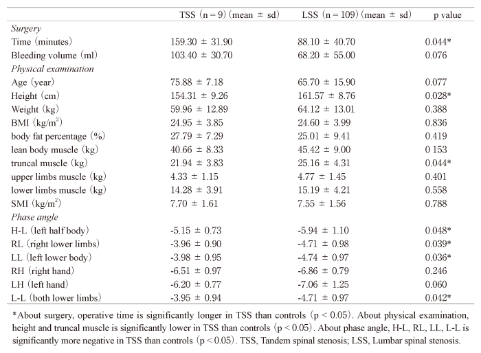
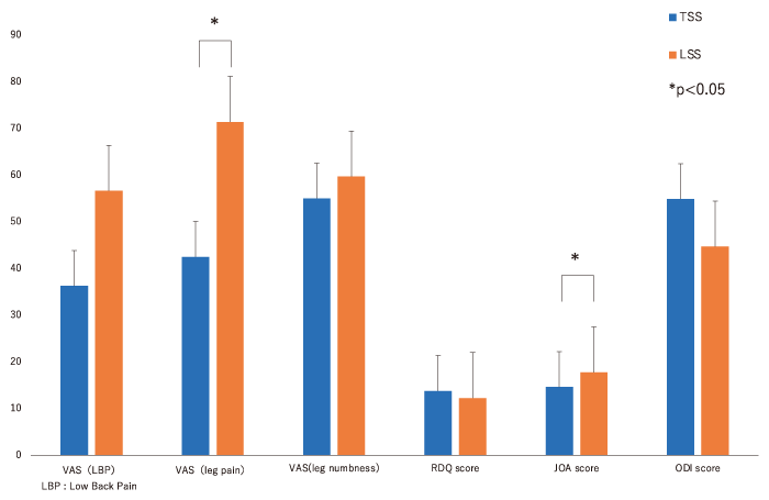
Fig. 1 Preoperative clinical symptoms in each group. VAS score (leg pain) and JOA score were significantly higher in LSS than in TSS (p = 0.007 and 0.016, respectively). VAS, visual analogue scale; VAS (LBP) : VAS (Low Back Pain) ; JOA, Japanese Orthopaedic Association; RDQ, Roland-Morris Disability Questionnaire; ODI, Oswestry Disability Index; TSS, Tandem spinal stenosis; LSS, lumbar spinal canal stenosis.
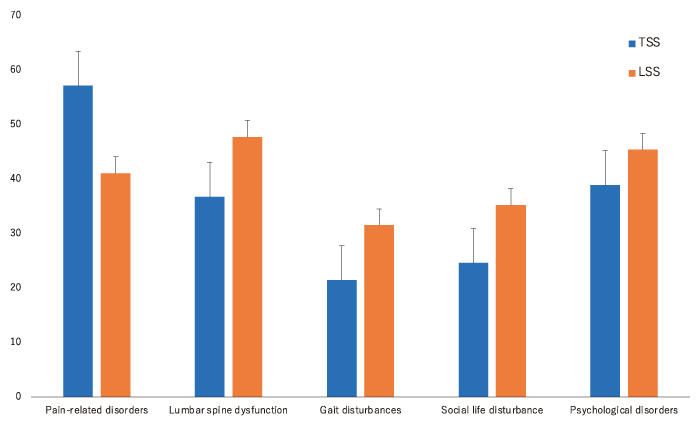
Fig. 2 Preoperative JOABPEQ scores in each group. There was no significant difference between the two groups in any category of the JOABPEQ. JOABPEQ, Japanese Orthopaedic Association Back Pain Evaluation Questionnaire; TSS, Tandem spinal stenosis; LSS, lumbar spinal canal stenosis.
Table 2 Percentage of cases in the 2 groups reporting symptoms based on the JOABPEQ
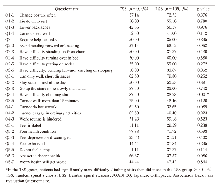
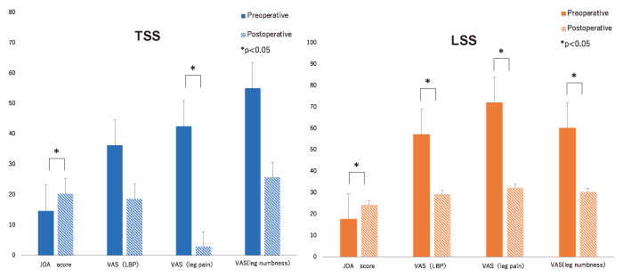
Fig. 3 Clinical symptoms before and after surgery in both groups. In the TSS group, JOA score and VAS (leg pain) were significantly improved (p = 0.028 and 0.021, respectively). On the other hand, there was significant improvement in all clinical measures in the LSS group (p < 0.001). TSS, Tandem spinal stenosis; LSS, Lumbar spinal stenosis; JOA, Japanese Orthopaedic Association; VAS, visual analog scale.
The pathophysiology and cause of TSS have not been clarified, and there is no consensus on the surgical method to treat it. In this study, we compared skeletal muscle mass, clinical symptoms, and surgical results between the TSS and LSS groups. Operative time was significantly longer in the TSS group than in the LSS group, but the amount of bleeding was not significantly different between the two groups. The trunk muscle mass and phase angle (left half body, right lower limb, left lower limb, both lower limbs) were significantly lower in the TSS group than in the LSS group, and about 90% of TSS cases were characterized by difficulty in climbing stairs. The present study revealed effectiveness of the one-stage surgery for TSS, which showed comparable clinical improvement as the surgeries for LSS alone with the same amount intraoperative bleeding.
The prevalence of TSS varies from report to report. Bajwa and Lee reported a prevalence of 2-5.4% based on a cadaver study[4,17]. Kawaguchi et al. reported that the prevalence of TSS was 19-60% when there was stenosis in either the lumbar region or the neck, using diagnostic imaging such as simple X-rays and CT[9].
Early suspicion of TSS is important because TSS often presents with residual symptoms and is often diagnosed only after surgery on either the cervical or lumbar spine.
Lee et al. showed lumbar spinal stenosis symptoms at multiple levels in men over 70 years old[4], indicating age and gender are risk factors. Iizuka et al. reported that the only other independent risk factor for TSS is a cervical Torg-Pavlov ratio (the ratio between the developmental sagittal diameter and the vertebral body diameter of the same level from cervical spine radiographs) of < 0.78 [12]. The Torg-Pavlov ratio is reported to be significantly lower in patients with cervical spondylotic myelopathy[18,19]. This is consistent with our observation that the TSS group had lower trunk muscle mass and lower phase angles of trunk and lower limb than the LSS group, and about 90% of cases had difficulty climbing stairs.
It is known that a decrease in trunk muscle mass greatly affects spinal alignment. Eguchi et al.[20] have shown that low trunk muscle mass is an ageindependent risk factor in lumbar degenerative scoliosis. Hori et al.[21]demonstrated that trunk muscle mass may signal lumbar spinal dysfunction and imbalance that may deteriorate when trunk muscle mass is less than approximately 23 kg. In this study, the trunk muscle mass was significantly lower in the TSS group, suggesting an effect of decreased activity.
The phase angle (PhA) is high in normal cells with a structurally complete cell membrane, as is found in healthy subjects and athletes. It is low in cells with structural damage to the cell membrane that occurs with aging, cancer and decreased cell density. Clinically known as an index for assessing nutritional status, low PhA is said to be significantly associated with nutritional risk, length of hospital stay, and mortality[22,23]. In recent years, the relationship between PhA and skeletal muscle mass also has attracted attention. In this study, PhA in the left half of the body and both lower limbs was significantly lower in the TSS group, suggesting that aging of the cell membranes in the tissues of the trunkand both lower limbs is progressive in TSS. In lumbar spinal stenosis, multi-level stenosis that involves the lower lumbar spine is more prone to severe neuropathy than single intervertebral stenosis. Spinal nerve compression in both the cervical and lumbar spine exacerbates the senescence of the trunk and lower limb skeletal muscle cells distal to the stenosis[24,25]. Detailed research on the relationship between PhA and spinal disorders is needed in the future.
The RDQ and Oswestry Disability Index are specialized scales for low-back pain related quality of life, but the SF-36 and EuroQol are widely used as comprehensive health assessments around the world. JOABPEQ is a patient-centered evaluation of treatment outcomes that takes into account both scientific and psychological factors[26]. An Excel file can be downloaded from the JOA website and used to automatically assess the severity of individual patients.
Using the JOABPEQ questionnaire in LSS patients, Eguchi et al. reported that resting leg pain, such as difficulty in wearing socks, is a characteristic of lumbar foraminal stenosis[27]. In this study, the proportion of gait dysfunction defined using the JOABPQ, specifically climb stairs, was higher in the TSS group than in the LSS group. This is consistent with the symptoms of TSS characterized by progressive gait disturbance[8]. If there is difficulty in climbing stairs that does not match the severity of LSS, it is necessary to treat TSS with cervical myelopathy in mind.
There is still no consensus on the surgical method for TSS, whether a one-stage approach or a twostage approach is preferable. Hsieh et al. showed that patients with symptomatic TSS who underwent cervical decompression and elimination of lumbar symptoms did not necessarily require tandem decompression[11]. Additionally, Epstein et al. found that all patients treated with primary cervical decompression had improvement in their lower extremity symptoms[1]. Recently, some surgeons have advocated for simultaneous decompression of both the cervical and the lumbar spine in patients with TSS. This approach utilizes two separate operative teams addressing the cervical and lumbar regions simultaneously. The simultaneous approach resulted in shorter operative time and less blood loss compared to staged surgery, although there was no difference in clinical outcomes[28-31]. In this study, one-stage surgery was performed on all TSS cases by a single surgical team and the amount of intraoperative bleeding was equivalent to that of LSS alone. Significant improvement in clinical symptoms was observed, suggesting that the one-team approach to TSS is effective.
Our study has several limitations. (1) Only a small number of subjects were investigated, requiring confirmation of our findings in a larger population. (2) The TSS group was older than the LSS group and may have had age-related loss of skeletal muscle mass. (3) One-stage surgery and staged surgery in the TSS group were not compared.
One-stage surgery for TSS is minimally invasive with intraoperative bleeding equivalent to that of LSS alone. Postoperative clinical results also were equivalent to those of LSS alone. A one-stage approach to TSS is considered effective.
YE conducted data collection and data entry, performed the statistical analysis, and wrote the manuscript. All authors contributed to and approved the final manuscript.
There is no financial supports.
We declare that there are no relevant conflicts of interest. Dr. Seiji Ohtori is a member of the journal’s editorial board.
The study protocol was approved by the ethics review committee (IRB approval no., H26’-4).
Available.
Address correspondence to Dr. Masaya Mizutani.
Department of Orthopaedic Surgery, Graduate School of Medicine, Chiba University,
1-8-1 Inohana, Chuou-Ku, Chiba 260-8670, Japan.
Phone: +81-43-222-7171.
Fax: +81-43-226-2116.
E-mail: anchan7117@gmail.com
