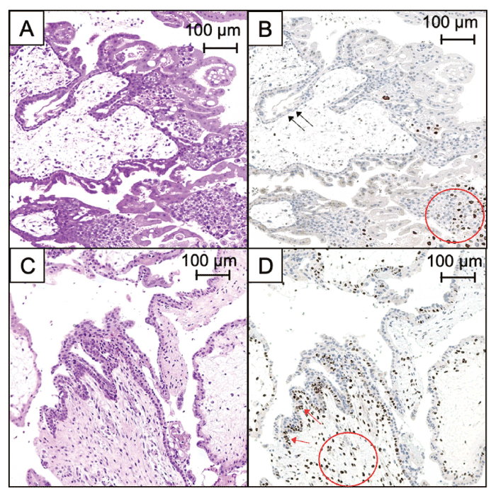
Fig. 1 Pathological features of hydatidiform moles. Complete hydatidiform moles (CHM). (A) Hematoxylineosin staining (× 100, scale bar: 100 μm); (B) immunohistochemistry of p57KIP2. Extravillous trophoblasts were stained (red circles) (× 100, scale bar: 100 μm). Cytotrophoblasts and stromal cells of CHM were not stained with anti-p57KIP2 antibody (black arrows). Partial hydatidiform moles (PHM). (C) Hematoxylin-eosin staining (× 100, scale bar: 100 μm). (D) Immunohistochemistry of p57KIP2 (× 100, scale bar: 100 μm). Cytotrophoblasts (red arrows) and stromal cells (red circles) of PHM were stained with anti-p57KIP2 antibody.