Fig. 1
Typical Images Acquired with 320 Slice Computed Tomography (CT) and by Invasive Coronary Angiography (ICA)of a Patient with Chronic Atrial Fibrillation (CAF)(. Modified from Reference 39).
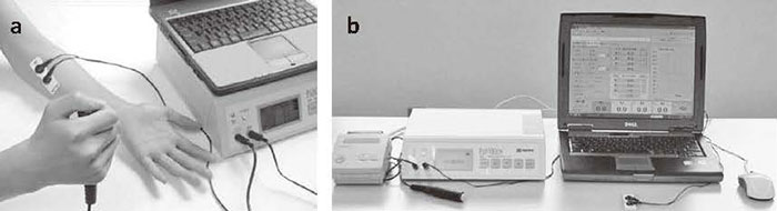
PainVision.( A) Electrodes are patched on the surface of patient forearms, and both current at perception threshold and current producing pain comparable with low back pain are measured.( B) The degree of pain is calculated automatically using software.