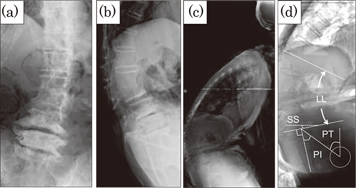
Fig. 2
Plain lumbar radiographs.( a) Anteroposterior, (b) Lateral( lumbar), and( c) Lateral( whole spine) images. The patient showed severe kyphoscoliosis.( d) Inversed image with sagittal parameters. LL: lumbar lordosis, SS: sacral slope, PT: pelvic tilt, PI: pelvic incidence( Further details are explained in the text.)