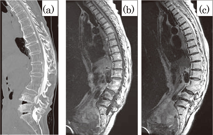
Fig. 3
(a)Computed tomography showed posterior components of the thoracic spine showed ankylosing changes.(b, c) Sagittal magnetic resonance imaging.(b) T1-weighed image(WI)(c) T2-WI. Fused vertebrae of L2-3 showed no septic intensity change. No findings related to active tuberculosis were evident.