


Chiba Medical J. 91E:19~27,2015
doi:10.20776/S03035476-91E-4-P19
[Review Article]
Junichi Nakamura, Seiji Ohtori, Sumihisa Orita, Shuichi Miyamoto,
Yasushi Wako, Michiaki Miura and Kazuhisa Takahashi
1) Department of Orthopaedic Surgery, Graduate School of Medicine, Chiba University, Chiba 260-8670.
(Received March 9, 2015, Accepted April 1, 2015)
Magnetic Resonance Imaging (MRI) was first utilized for the diagnosis of osteonecrosis in 1983.
The purpose of this current study is to review the past 30 years of achievement using new developments in MRI technology for investigation into the pathogenesis of osteonecrosis of the femoral head associated with corticosteroid therapy. From 1983 to 2013, 165 MRI studies were reported: 25 articles in 1983-1993, 55 in 1994-2003, and 85 in 2004-2013. Forty-nine articles were reported from Japan, 44 from the United States, and 18 from China. Eighty-two studies were retrospective, 49 were prospective, 20 were case reports, two were genotyping studies, and 10 used animal models. MRI is a promising tool for monitoring osteonecrosis and analyzing the pathogenesis in both clinical and experimental studies.
Review, Magnetic resonance imaging, Corticosteroid, Osteonecrosis of the femoral head
Osteonecrosis of the femoral head impairs quality of life in young and active individuals [1]. Corticosteroidassociated osteonecrosis in systemic lupus erythematosus (SLE) was first reported by Dubois and Cozen in 1960[2]. The pathogenesis of osteonecrosis has been unclear to date, but corticosteroid use has been widely accepted to be influential in the development of osteonecrosis [3]. Corticosteroid therapy is an ideal way to reveal the pathogenesis of osteonecrosis because it is straightforward to evaluate the precise dosage and duration of corticosteroid therapy in patients with osteonecrosis.
Magnetic Resonance Imaging (MRI) was first utilized for the diagnosis of osteonecrosis in 1983 [4,5]and is now the gold standard for initial diagnosis of osteonecrosis because MRI is more sensitive in demarcating normal bone from the necrotic lesion much earlier than simple X-rays. The purpose of this study is to review the improvements that the development of MRI technology has made in the investigation into the pathogenesis of osteonecrosis of the femoral head associated with corticosteroid therapy 30 years after the first clinical application of this imaging methodology.
A systematic review was performed using PubMed (US National Library of Medicine, National Institutes of Health) on January 1st 2014. Inclusion criteria were original articles written in English and published between 1983 and 2013 related to corticosteroidassociated osteonecrosis of the femoral head using MRI.
Exclusion criteria were review articles or other unrelated papers. Filters for key words were“ osteonecrosis” AND (“femoral head” OR“ hip”) AND“ MRI” AND (“steroid” OR“ steroids” OR“ corticosteroid” OR“ corticosteroids”) AND“ English [lang]”. Articles during this 30 year period were divided into three groups by 10 year increments: 1983-1993, 1994-2003, and 2004- 2013. The number of articles, country of contribution, type of study, and impact factor in 2013 (Journal of Citation Reports, Thomson Reuters, New York) were analyzed. Furthermore, MRI-based etiology, diagnosis, classification, prognosis, and treatment outcome were reviewed.
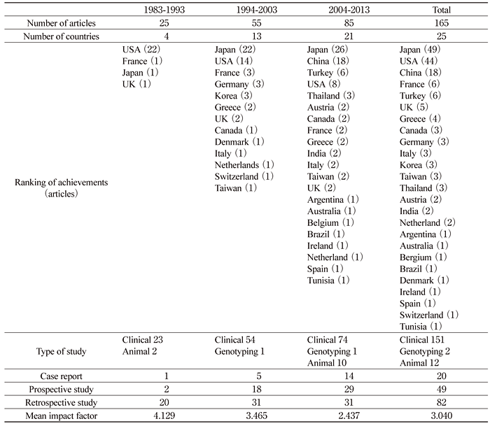
We identified 203 articles via PubMed published between 1983-2013 of which 165 met the inclusion criteria. The number of MRI studies increased each decade: 25 articles in 1983-1993, 55 in 1994-2003, and 85 in 2004-2013 (Table 1). Forty-nine articles were reported from Japan, 44 from the United States, and 18 from China. In Japan, 16 of 49 articles (33%) were studied in Chiba University. Reports were received from four countries in 1983-1993, 13 countries in 1994- 2003, and 21 countries in 2004-2013. However, over 30 years, 67% of the articles about osteonecrosis generated were from Japan, the U.S. or China. In the last decade, Japan and China in particular increased the number of their publications in this area. Clinical studies comprised 151 articles: 82 were retrospective studies, 49 were prospective studies, and 20 were case reports. MRI prospective studies increased over the three decades. In the last decade, experimental studies using MRI were identified in two articles about genotyping and in 10 articles using animal models. The mean impact factor in 2013 of the journals that published these articles was 3.04 (range: 0 to 17.879).
Table 1 MRI articles of osteonecrosis of the femoral head by decade
The phenomenon of nuclear magnetic resonance was discovered in 1946[6,7], and it was demonstrated as the basis of an imaging technique in 1973[8]. The appearance of MRI made it possible by 1983 to visualize lesions of the musculoskeletal system[4,5]. MRI has rapidly become the gold standard for early diagnosis of osteonecrosis because of its compatibility with histology [9]. MRI is a more sensitive and reliable technique than conventional X-rays, computed tomography or radionuclide bone scanning, with 85-100% sensitivity and 98% specificity for the early diagnosis of osteonecrosis of the femoral head[10-12]. Immediately after ischemia, the ischemic area of bone marrow is not visible by MRI. However, the healing and growth of the fibrous reactive tissue into the necrotic area replaces the dead marrow, and can be identified with MRI by its different characteristics. A low-signal intensity band represents the repair tissue interface surrounding a high-signal-intensity necrotic marrow segment. MRI and single-photon emission computed tomography can detect osteonecrosis within 4 months after renal transplantation[13], and can be used to monitor the course of the affected hip in patients with osteonecrosis [14]. Characteristic abnormalities of osteonecrosis on MRI are described by patterns that are homogeneous, inhomogeneous, ring, or band patterns[15]. The early conversion from hematopoietic marrow to fatty marrow in patients with osteonecrosis of the femoral head may reflect decreased vascularity of the proximal femur [16]. Cova et al [17]. developed a dog model to evaluate bone marrow perfusion using gadolinium-enhanced dynamic MRI and probed the peak percentage of enhancement (i.e. blood supply) using blood-borne microspheres. In another dog model, dynamic contrast-enhanced MR imaging proved significantly more sensitive than spin-echo and short tau inversion recovery imaging in the earlier detection of acute avascular necrosis [18]. Normal marrow was characterized by rapid enhancement, although the marrow of the ischemic femoral head with avascular marrow showed persistent lack of enhancement. A junctional zone, characterized by rapid contrast enhancement in excess of 120% without early washout, was identified at the interface between normal and avascular marrow. Kopecky et al [19]. reported the first MRI prospective study for 24 months after renal transplantation in 104 recipients, with 12% incidence of osteonecrosis of the femoral head. Of 25 affected hips, seven hips became painful but 18 hips remained asymptomatic. All MR lesion sizes in the symptomatic hips were larger than those in the asymptomatic hips. MR lesions regressed in size in seven hips and disappeared in six hips, showing spontaneous improvement.
Corticosteroid-associated osteonecrosis is multifocal, with 66% of the MRI-identified lesions in the hip, 51% in the knee, 16% in the ankle, and 16% in the shoulder [20]. Bilateral MRI screening of the hip and knee is an effective method to diagnose osteonecrosis in patients with high dose corticosteroid therapy (more than 30mg/ day). The incidence of osteonecrosis of the femoral head has been reported in prospective observational MRI studies as 32% in various autoimmune-related disorders[21], 44% in SLE patients[22], and 23% in renal allograft recipients[23]. Moreover, osteonecrotic lesions were identified 3-4 months after initiation of corticosteroid therapy in those studies[21-23]. The incidence of osteonecrosis was much higher and the development of osteonecrosis much earlier than expected based on conventional X-rays because MRI can detect asymptomatic and non-collapsed osteonecrosis. Therefore, an ischemic event that causes osteonecrosis seems to occur within 3-4 months after corticosteroid administration, considering the time lag of the reparative reaction to the dead bone. The initial MR sign of asymptomatic osteonecrosis in the early stage is a band-like pattern; bone marrow edema is a sign of collapse with advanced stage osteonecrosis that occurs with pain[23,24]. Bone marrow edema is easily detected with fat suppression images[25], but this makes it difficult to distinguish the viable area from the necrotic area. Gadolinium-enhanced MRI can demarcate the boundary of the reactive zone[26].
Lesion size and lesion location are both strongly correlated with risk of collapse in osteonecrosis[27]. The survival rate at 32 months was 100% for the small osteonecrotic lesions within one-third of the weightbearing area of the acetabulum in coronal MRI (Grade a)[27]. The survival rate was 86% for the moderate lesions between one-third and two-thirds of the weightbearing area (Grade b), and 29% for the large lesions, which encompassed more than two-thirds of the weightbearing area (Grade c)[27]. Kubo et al. [23]found same evidence that a large lesion (Grade c) became symptomatic 7-14 months after renal transplantation and then progressed to collapse. Based on these facts, the Japanese Ministry of Health, Labor and Welfare (JMHLW) established a classification using the boundary of a necrotic lesion as a low-intensity band on the central coronal section of the femoral head on T1-weighted images[28]. Lesions are classified into the following four types: type A-lesion occupies the medial one-third or less of the weight-bearing area; type B-lesion occupies the medial two-thirds or less of the weight-bearing area; type C1-lesion occupies more than the medial two-thirds of the weight-bearing area but does not extend laterally to the acetabular edge; and type C2-lesion extends laterally to the acetabular edge. The MRI-based JMHLW type classification showed excellent inter-observer and intra-observer reliabilities (weighted kappa of 0.709-0.724 and 0.780-0.800, respectively) [29]. Other methods have been used to attempt to quantify the extent of osteonecrosis. Lafforgue et al [30]. proposed three quantitative parameters: the angle filled by the lesion (alpha); the percentage of weightbearing femoral cortex that was osteonecrotic; and the percentage of osteonecrotic femoral head surface. Steinberg et al [31,32]. calculated the percentage of lesion size by estimating the average area of abnormality on serial MR images, classifying them into three groups based on lesion size: Group A, less than 15% of femoral head involvement; Group B, 15% to 30%; and Group C, greater than 30%. Koo et al [33]. developed a formula from a combination of the arc of the necrotic portion in the mid-coronal image (A) and that in the mid-sagittal image (B): (A/180)×(B/180)×100. Nishii et al [34]. performed a three-dimensional quantification of lesion volume as well as latitude and longitude of the center of gravity of the lesion within the femoral head.
Aggressive treatment such as core-decompression is undertaken for asymptomatic osteonecrosis [14-16,30-33], presumably because spontaneous healing or regression of the lesion has been believed to be extremely rare[35]. However, Sakamoto et al [21]. observed spontaneous reduction of lesion size within one year after corticosteroid therapy in 45% of osteonecrosis cases, although there was no further change with a longer follow-up of 31 months. Oinuma et al [22]. reported that no new lesions were detectable from 6 months to 12 months after corticosteroid therapy in SLE patients. Kubo et al. also reported that no new abnormal findings on MRI were detected between one year and 4.3 years in renal allograft recipients[23]. Osteonecrosis after renal transplantation has decreased because the development of novel immunosuppressant agents has decreased the necessity for corticosteroid treatment [36]. Pritchett et al [37]. suggested that statins have a preventive effect against developing osteonecrosis, observing that treatment reduced the incidence of osteonecrosis to only 1% in patients who received statin drugs during the entire time of steroid exposure. High-dose corticosteroid treatment causes hemostatic abnormality. Plasmin-α2-plasmin inhibitor complex was significantly higher in autoimmune disease patients with osteonecrosis than in those without [38]. The number of osteonecrotic joints was correlated with the plasmin inhibitor levels. Hypofibrinolysis conferred by the 4G/4G plasminogen activator inhibitor-1 gene variant is a major predisposing factor for osteonecrosis in renal transplant recipients [39].
The pathogenesis of corticosteroid-associated osteonecrosis is still controversial, but the dosage of daily oral corticosteroids seems the most influential factor. A high dose of oral corticosteroids (>40mg/ day) is a risk factor for development of osteonecrosis [40], partly because blood supply to the femoral head decreases in the early period after high-dose corticosteroid therapy. Blood supply can increase when the corticosteroid dose is reduced[41]. De novo osteonecrosis develops after increased corticosteroid use for SLE recurrence in patients who did not have osteonecrosis for at least ten years prior to the induction of corticosteroid use[42,43]. Other risk factors for osteonecrosis have been hypothesized, such as age, underlying disease, sex, and alcohol. Dynamic enhanced MRI reveals that blood supply to the femoral head is more abundant in children (especially at the growth plate) than in adults[41]. This characteristic seems to explain the lower incidence of osteonecrosis in children [44,45]. Pediatric SLE patients showed a lower rate of osteonecrosis than adolescent and adult patients (6% versus 41%)[44]. In pediatric acute lymphoblastic leukemia, the median age at corticosteroid induction was 13.5 years for patients who developed osteonecrosis and was 4.7 years for those without osteonecrosis [45]. Corticosteroid-associated osteonecrosis has been reported in various disorders, but the incidence of osteonecrosis in SLE patients is significantly higher than in those with other autoimmune diseases (37% versus 21%)[40]. SLE patients have a significantly higher risk of osteonecrosis than non-SLE patients (Odds ratio= 2.6), and male patients have a significantly higher risk than female patients (Odds ratio=1.6). Alcohol intake is also a risk factor for developing osteonecrosis, but the pathogenesis seems to be slightly different from that of corticosteroid-induced osteonecrosis, and is specific to the femoral head. The incidence of alcohol-associated osteonecrosis of the knee is lower than the incidence of steroid-associated osteonecrosis of the knee in patients with osteonecrosis of the femoral head[46]. Vande Berg et al [47]. proposed that baseline femoral neck status was a predictive factor for developing corticosteroidassociated osteonecrosis of the femoral head because osteonecrosis was correlated with high marrow fat in the proximal femur before corticosteroid therapy, supporting the bone marrow conversion theory of Mitchel et al [16].
The natural history of osteonecrosis of the femoral head has been monitored longitudinally with MRI. The frequency of articular collapse was significantly higher in osteonecrosis of the femoral head than in osteonecrosis of the knee (38% versus 5%)[48]. After articular collapse, surgery was performed significantly more often for osteonecrosis of the femoral head than for osteonecrosis of the knee (63% versus 29%)[48]. On the other hand, spontaneous repair of osteonecrosis has been revealed by long-term observational MRI studies [49-53]. Spontaneous repair of the lesion occurred in 61% of SLE patients over 10 years of follow-up[49]. The lesion volume was reduced in 95% of patients with severe acute respiratory syndrome who developed osteonecrosis of the femoral head, but the remaining hips with relatively larger lesion volumes showed no apparent reductions over five years of follow-up[52]. The reduction in lesion size observed on MRI is a slow, discontinuous and time-dependent process. Factors favorable to resolution are early diagnosis, asymptomatic disease, and small lesion size[50].
Other novel imaging techniques also have been developed to assess changes associated with osteonecrosis. T2 mapping, a quantitative evaluation of cartilage matrix status including hydration and collagen fiber integrity, revealed articular cartilage degeneration with non-collapsed and asymptomatic osteonecrosis of the femoral head in SLE patients[54]. Moreover, acetabular dysplasia was associated with high T2 values. Positron emission tomography showed increased uptake of fluoride for glucose in the acetabulum in 9 of 17 hips with osteonecrosis of the femoral head, reflecting inflammation and degeneration[55]. Whole body MRI may become the ideal imaging modality of choice in the future, especially for patients receiving high dose corticosteroids who are at high risk of developing osteonecrosis at multiple sites [56,57].
MRI based experimental studies also have been conducted. A rabbit model of corticosteroid-induced osteonecrosis of the femoral head was developed in which MRI detected bone marrow edema and spotlike high signals in T2-weighted images of cancellous bone[58]. Dynamic MR imaging of the rabbit model showed a significantly decreased peak percentage of enhancement (i.e., blood supply) at the proximal femur with osteonecrosis compared with controls without osteonecrosis[59]. MRI screening of corticosteroidinduced osteonecrosis of the femoral head in the rabbit model was utilized to investigate the efficacy of the core decompression treatment [60]. Decompression took partial effect in promoting bone regeneration in the early stage, but the long-term effect was not satisfactory. Granulocyte colony-stimulating factor and stem cell factor promoted new vessel formation and new bone formation in corticosteroid-associated osteonecrosis in rabbits[61]. As for another animal model, Zheng et al [62]. reported a bipedal emu model of corticosteroidinduced osteonecrosis of the femoral head that could be detected by MRI. Gene expression profiling of single nucleotide polymorphisms has shown that cAMPresponsive element binding protein-binding proteins may affect susceptibility to steroid-induced osteonecrosis in renal allograft recipients [63]. Another study suggests that apolipoprotein B C7623T polymorphisms can predict the risk for osteonecrosis before corticosteroid administration[64]. In conclusion, MRI is a promising tool for monitoring osteonecrosis and analyzing its pathogenesis in both clinical and experimental studies.
The corresponding author, JN, received Grants-in- Aid for Scientific Research (Research Project Number: 25870125) for this work. The other authors do not receive any funding or financial support that may be perceived to have biased the study.
Address correspondence to Dr. Junichi Nakamura.
Department of Orthopaedic Surgery, Graduate School of Medicine, Chiba University, 1-8-1, Inohana, Chuou-ku, Chiba, 260-8670 Japan.
Phone: +81-43-226-2117. Fax: +81-43-226-2116.
E-mail: njonedr@chiba-u.jp
Abbreviations: Systemic lupus erythematosus (SLE), Magnetic Resonance Imaging (MRI)
