


Chiba Medical J. 92E:31~36,2016
doi:10.20776/S03035476-92E-4-P31
[ Original Paper ]
Takanori Omae, Junichi Nakamura, Seiji Ohtori, Sumihisa Orita
Takane Suzuki, Miyako Suzuki, Shuichi Miyamoto, Shigeo Hagiwara
Takayuki Nakajima, Makoto Takazawa, Tomonori Shigemura
Yasushi Wako, Michiaki Miura, Yuya Kawarai, Masahiko Sugano
Kento Nawata and Kazuhisa Takahashi
Department of Orthopedic Surgery, Graduate School of Medicine, Chiba University, Chiba 260-8670.
(Received March 14, 2016, Accepted March 23, 2016)
Objectives To determine gait and inflammatory response in a rat hip pain model produced by intra-articular injection of nerve growth factor(NGF).
Methods Using 36 8-week-old male Sprague Dawley rats, 30 µl of saline(sham-operated group; n=12), 30 µL of 50 µg/mL of NGF(NGF50 group; n=12), and 30 µL of 100 µg/mL of NGF(NGF100 group; n=12) were injected into the left hip. The gait of 6 rats in each group was analyzed using a CatWalk system. The levels of inflammatory cytokines were determined with the synovium and femoral head.
Results The maximum contact area was significantly decreased in the NGF100 group compared with in the sham-operated group on day 14. Swing speed significantly decreased in the NGF50 and NGF100 groups compared with in the sham-operated group on day 14. Duty cycle significantly decreased in the NGF50 and NGF100 groups compared with the sham-operated group on day 14. TNF-α, IL-1β, and IL-6 were significantly elevated in NGF50 and NGF100 groups compared with the sham-operated group.
Conclusions NGF into the rat hip joint induces an increase of inflammatory cytokines in the synovium and elicits pain escape behavior.
Gait, cytokine, hip joint, nerve growth factor
The pathogenesis of hip pain is unclear and the roles of sensory innervation and the nature of pain transmitting substances in the hip joint have not been fully elucidated. Previously, we reported that dorsal root ganglion(DRG) neurons innervating the hip are distributed on multiple levels(L1-L4) [1], and that there is a difference in the level of innervation between the hip and inguinal skin[2]. Moreover, DRG neurons that innervate the hip and inguinal skin may overlap [3]. Animal models of hip pain are essential to clarify the pathogenesis of hip pain. However, such models of hip pain have not been widely reported. We have established two novel rat models of hip pain produced by intra-articular administration into the hip joint with mono-iodoacetate [4]or nerve growth factor(NGF) [5]. NGF is a neurotrophin that is well known to regulate neuronal development, survival, and maintenance [6]. NGF also seems to influence chronic pain states, notably those associated with inflammation, and may be a target for analgesia in patients with osteoarthritis(OA) [7]. Intra-articular injection of NGF into rat hips produced microinflammation without osteoarthritic change [5]. Intra-articular injection of NGF into rat knees evoked pain-related behavior [8]. NGF is associated with angiogenesis at the osteochondral junction in patients with arthritis [9]. We hypothesized that direct application of NGF into the hip joint evokes an inflammatory condition without degeneration of the articular cartilage and that NGF can be used to model inflammatory hip pain in rats. However, the direct effects of intra-articular injection of NGF into normal rat hips on pain mediators and pain-related behavior are not yet known.
A CatWalk gait analysis system is able to perform detailed gait analysis noninvasively and repeatedly [10]. This system has been described in detail elsewhere [11-14]. The time course of pain-related mediator appearance can be analyzed using enzyme-linked immunosorbent assay(ELISA) to determine the levels of inflammatory cytokines, focusing on inflammatory and neuropathic pain-related states using local tissues and peripheral sensory innervation including DRGs.
The purpose of this study was to evaluate painrelated behavior using the CatWalk gait analysis system and the synovial levels of inflammatory cytokines in a rat model of hip pain produced by intra-articular injection of NGF into the hip joint.
We used 36 8-week-old male Sprague Dawley rats weighing from 250 to 300 g. The protocols for the animal procedures in our experiments followed the 1996 revision of the National Institutes of Health guidelines for the Care and Use of Laboratory Animals and received prior approval from the ethics committee of our institution. The rats were anesthetized by an intraperitoneal injection of sodium pentobarbital(40mg/kg) and were treated aseptically throughout the experiments. Using a 26-gauge needle, the following solutions were injected into the left hip of rats assigned to various groups according to their treatment: 30 µL of saline in rats in a sham-operated group(n=12), 30µL of 50 µg/mL of NGF(recombinant human b-NGF, 256-GF-100/CF; R&D Systems, Minneapolis, MN) in rats in a NGF50 group(n=12), and 30 µL of 100µg/ mL of NGF in rats in a NGF100 group(n=12). The intra-articular injections were limited to the joint cavity. The rats were allowed unrestricted cage activity after the injection.
The CatWalk system (Noldus Information Technology, Wageningen, The Netherlands) was used to evaluate gait characteristics in the present study. Rats were placed on a glass plate located in a darkened room and allowed to walk freely. A light beam from a fluorescent lamp was shone through the glass plate. The light was normally reflected internally, except in areas where the rat’s paw touched the glass plate, where the light was refracted on the opposite side. This produced a sharp bright image of the paw print as seen from under the plate where the rat walked, and the entire run was recorded using a video camera. The data were acquired, compressed, and analyzed using CatWalk system software. In the present study, the gait patterns of 6 rats in each group were recorded 3 times and analyzed automatically using the CatWalk system.
We compared 18 parameters of hind paw gait between the 3 groups. The ratios of the affected side to the unaffected side were taken as adjusted data for each parameter. The rat gait patterns were recorded on day 0(before injection, n=6) and again on days 7 and 14 after the injection(before they were humanely killed, n=6, respectively)
For each group, 6 rats were killed after Experiment 1 on day 7, while the remaining 6 were killed on day 14. Rats were deeply anesthetized using 40mg/kg sodium pentobarbital and their left hips were exposed; the synovium and femoral head cartilage were resected for cytokine ELISAs. Tumor necrosis factor(TNF)-α, interleukin (IL)-1β, and IL-6 production in the synovium and the femoral head cartilage were quantified using ELISAs in accordance with the manufacturer’s protocols(R&D Systems, Minneapolis, MN). Tissue protein was assayed using a kit in accordance with the manufacturer’s protocol(Bio-Rad, Hercules, CA), and cytokine levels were normalized to tissue protein levels.
All values were calculated and statistically compared using unpaired t test followed by Bonferroni’s correction. A P<0.05 was considered to be significant.
Of 18 parameters for hind paws, maximum contact area(the maximum area of paw contact with the glass plate), swing speed(the speed of the paw during gait), and duty cycle(duration of paw contact divided by time between consecutive paw contacts: contact(/ contact+ swing)×100%) were determined to be significantly different between the 3 groups. Maximum contact area tended to decrease from day 0 to day 7 after NGF injection in rats in all 3 groups and then significantly decreased in rats in the NGF100 group compared with rats in the sham-operated group on day 14(81.3% versus 95.8%, P=0.0002, Figure 1). Swing speed tended to decrease from day 0 to day 7 after NGF injection in rats in all 3 groups and then significantly decreased in rats in both the NGF50 and NGF100 groups compared with rats in the sham-operated group on day 14(80.3% versus 82.2% versus 96.2%, P= 0.0001 and P=0.0005, respectively, Figure 2). Duty cycle tended to decrease from day 0 to day 7 after NGF injection in rats in all 3 groups and then significantly decreased in rats in both the NGF50 and NGF100 groups compared with rats in the sham-operated group on day 14(82.4% versus 83.0% versus 96.7%, P= 0.0007 and P=0.0005, respectively, Figure 3)
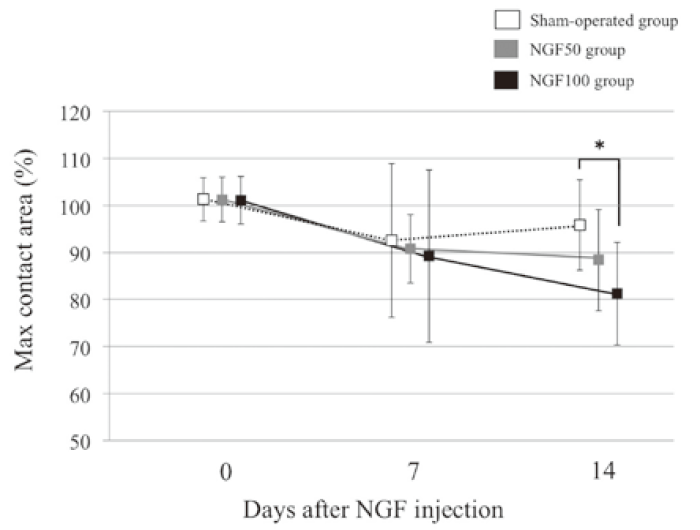
Fig.1
Maximum contact area.
The values are adjusted by the ratio of the affected to unaffected side (%). Maximum contact area was significantly decreased in rats in the NGF100 group compared with rats in the sham-operated group on day 14. Bars represent standard deviation. *P<0.05.
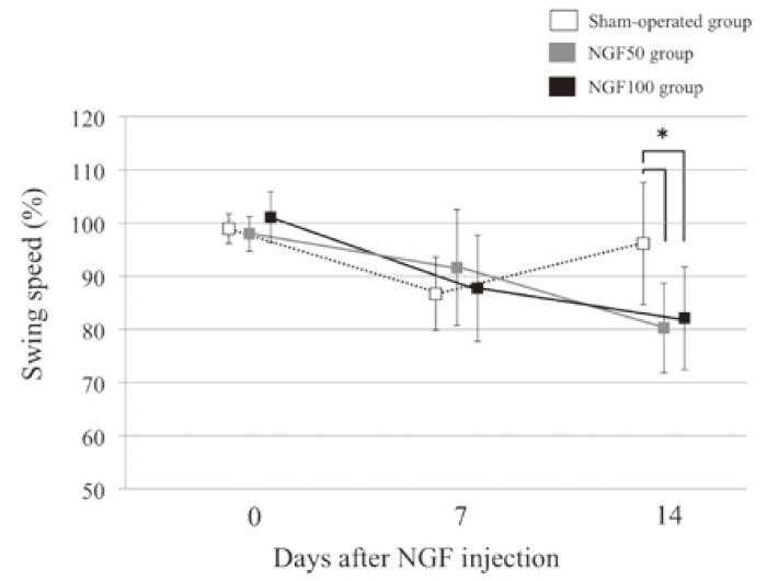
Fig.2
Swing speed.
The values are adjusted by the ratio of the affected to unaffected side(%). Swing speed was significantly decreased in rats in both the NGF50 and NGF100 groups compared with that in rats in the sham-operated group on day 14. Bars represent standard deviation. *P<0.05.
Synovial membrane levels of TNF-α (pg/mg protein) were significantly elevated on day 14 after the injection compared with day 7 in rats in both the NGF50 and NGF100 groups(1.46 versus 1.08, P=0.0019 and 1.55 versus 1.12, P=0.0004, respectively, Figure 4). The TNF-α levels were also significantly higher in rats in the NGF100 group than in rats in the sham-operated group on day 14 (1.55 versus 1.22, P=0.0025). Synovial membrane levels of IL-1β(pg/mg protein) were significantly higher in rats in both the NGF100 and NGF50 groups than in rats in the sham-operated group on day 7(11.2 versus 9.8 versus 7.8, P=0.0001 and P=0.0006, respectively, Figure 5). However, the difference was no longer significant on day 14. Synovial membrane levels of IL-6 (pg/mg protein) were significantly higher in rats in the NGF100 group than in rats in both the sham-operated and NGF50 groups on day 14(39.4 versus 33.1 versus 30.8, P=0.0007 and P=0.0001, respectively, Figure 6), although no significant difference was found on day 7.
There were no significant differences in cytokine levels in the femoral head cartilage at any time after the various treatments. The TNF-α levels in the femoral head cartilage were 1.22, 1.21, and 1.23 pg/mg protein in rats in the sham-operated, NGF50, and NGF100 groups on day 7, and 1.25, 1.27, and 1.29 pg/mg protein on day 14, respectively. IL-1β levels in the femoral head cartilage were 23.7, 24.6, and 24.9 pg/mg protein in rats in the sham-operated, NGF50, and NGF100 groups on day 7, and 23.8, 26.3, and 27.3 pg/mg protein on day 14, respectively. IL-6 levels in the femoral head cartilage were 106.1, 112.3, and 116.8 pg/mg protein in rats in the sham-operated, NGF50, and NGF100 groups on day 7, and 105.4, 105.6, and 97.7 pg/mg protein on day 14, respectively.
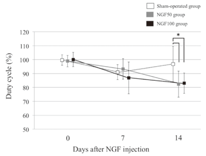
Fig.3
Duty cycle.
The values are adjusted by the ratio of the affected to unaffected side(%). Duty cycle was significantly decreased in rats in both the NGF50 and NGF100 groups compared with that in rats in the sham-operated group on day 14. Bars represent standard deviation. *P<0.05.
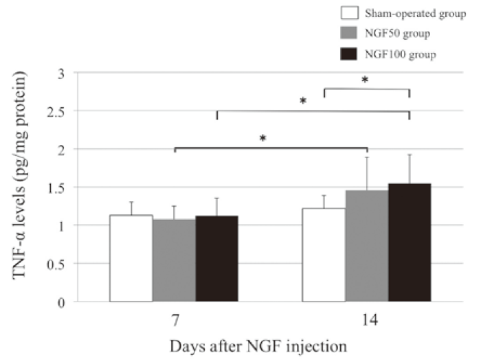
Fig.4
Synovial levels of TNF-α.
The synovial levels of TNF-α were significantly elevated on day 14 after the intra-articular injection of NGF compared with levels on day 7 in rats in both the NGF50 and NGF100 groups. The synovial levels of TNF-α were significantly higher in rats in NGF100 group than in rats in the shamoperated group on day 14. Bars represent standard deviation. *P<0.05.
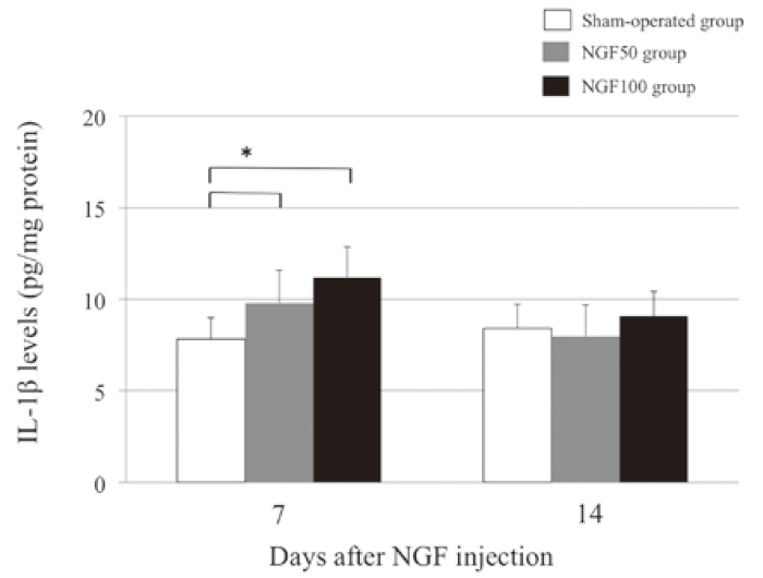
Fig.5
Synovial levels of IL-1β.
The synovial levels of IL-1β were significantly higher in rats in the NGF100 group than in rats in the shamoperated group on day 7. Bars represent standard deviation. *P<0.05.
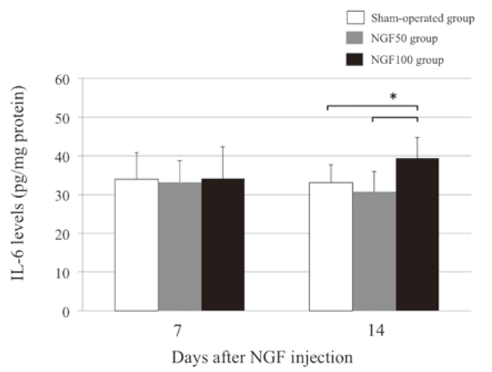
Fig.6
Synovial levels of IL-6.
The synovial levels of IL-6 were significantly higher in rats in the NGF100 group than in rats in both the shamoperated and NGF50 groups on day 14. Bars represent standard deviation. *P<0.05, **P<0.01.
To our knowledge, this is the first report describing hip pain-associated changes in gait parameters determined by CatWalk, including maximum contact area, swing speed, and duty cycle. Intra-articular injection of NGF into the hip joint apparently elicits pain escape behavior in rats. Miyagi et al. reported a rat model of myofascial inflammation using painassociated changes of gait parameters assessed using the CatWalk system [11]. In the model of myofascial inflammation, duty cycle of front and hind paws was significantly greater, stride length(the distance between successive placements of the same paw) of each paw was significantly shorter, and minimum contact intensity of the complete paw and mean contact intensity of each paw in the myofascial inflammation group were significantly greater compared with the control group. Yamazaki et al. reported a rat model of unstabilized rotator cuff defect using pain-associated changes of gait parameters assessed using the CatWalk system [13]. In the model of unstabilized rotator cuff defect, stride length, print area(the area of the paw contact with the glass runway surface during locomotion), and contact intensity(the mean intensity of all the pixels contributing to the maximum paw print area) were significantly less than in the control group.
The current report is also the first describing a rat model of hip pain associated with an increase in synovial levels of proinflammatory cytokines, including TNF-α, IL-1β, and IL-6 levels as assessed by ELISA. Intra-articular NGF administration into the rat hip was associated with an increase in the synovial level of inflammatory cytokines. Sensory innervation and inflammatory cytokines in hypertrophic synovial membranes are associated with nociception in OA of the hip in humans [15]. IL-6 and TNF-α levels are elevated in synovial joint fluid in patients with hip disease, reflecting tissue destruction in OA [16]. In the present study, synovial levels of TNF-α were significantly elevated on day 14 after the injection compared with the levels on day 7 in rats in both the NGF 50 and 100 groups. Synovial levels of IL-1β were significantly higher in rats in both the NGF 50 and 100 groups than in rats in the sham-operated group on day 7, although the difference was no longer significant on day 14. IL-1β is therefore likely to play a role in early inflammation. Synovial levels of IL-6 were significantly higher in rats in the NGF 100 group than in rats in the sham-operated and NGF 50 groups on day 14, although the levels were not significantly different on day 7. Thus, IL-6 is likely to play a role in delayed inflammation. Our findings suggest that NGF is crucially involved in the behavioral expression and neural transmission of pain from the hip joint.
There are several limitations to this study. First, dose dependency was not determined because only 2 doses of NGF were examined. Further doses of NGF should be studied to determine dose dependency. Second, gait parameters and inflammatory response were evaluated at only 2 time points. Pain-related behavior was significantly different between rats in the various treatment groups on day 14. In a previous study, pathology of the hip and immunohistochemistry of the DRG showed mild synovitis and expression of calcitonin gene-related peptide-immunoreactive neurons in sensory nerves on day 7 that persisted on day 14 [5] Determination of longer and shorter-term outcomes of intra-articular NGF injection is warranted. Third, in the CatWalk system, it is not possible to assess whether parameters are related to pain or muscle dysfunction without pain, and so the possibility that effects are related to muscle dysfunction cannot be excluded. In conclusion, an increase in synovial levels of inflammatory cytokines and pain-related escape behavior is observed in a novel rat model of hip pain induced by intra-articular administration of NGF into the hip joint.
Corresponding author, Junichi Nakamura received JSPS KAKENHI Grant Number 25870125.
Address correspondence to Dr. Junichi Nakamura.
Department of Orthopedic Surgery, Graduate School of Medicine, Chiba University, 1-8-1, Inohana, Chuou-ku, Chiba, 260-8670 Japan.
Phone: +81-43-226-2117.
Fax: +81-43-226-2116.
E-mail: njonedr@chiba-u.jp
Abbreviations:
dorsal root ganglion(DRG), nerve growth factor (NGF), osteoarthritis(OA), enzyme-linked immunosorbent assay(ELISA), tumor necrosis factor(TNF), interleukin(IL)
